Morphological Terms/Mesosoma
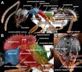
Aphaenogaster morphology. A: worker lateral; B: queen lateral, C: queen head. Provided by Minsoo Dong.

Terms use by Bolton (1994).


Abdomen
The classical third tagma of the insect body. The abdomen in ants consists of ten segments, of which the first seven (A1–A7, from front to back) are visible in the female castes (workers and queens), while A8 is also exposed in males. The tergites of segments A1–A8 each bears a spiracle, which may be exposed or concealed. In the female castes, segments A8 and A9 are desclerotised, internal, and form parts of the sting apparatus, so that A7, because it is always the last visible segment, is usually referred to as being apical. In males, A8 is exposed but A9 is usually retracted and is the gonosomite, having the male genitalia attached to its posterior margin. Segment A10 is reduced in both sexes, at most a simple tergal arc of cuticle. In males, tergite 10 is sometimes fused to tergite 9 (= syntergite), and in many groups the sclerite bears a pair of pygostyles (= cerci) apically.
The terminologies used to describe the ant abdomen may at first seem confusing. This is because two different systems tend to superimpose, and in places they are not strictly compatible.
- A terminology based strictly on morphology, which simply numbers the visible abdominal segments from front to back. This has the clear advantage of indicating homologous segments between different ant taxa, regardless of the specialisations of individual segments or groups of segments in different groups of ants.
- A more casual terminology, based on observed subdivisions of the abdominal segments, which names various specialised segments or groups of segments. The advantage here is that the subdivisions are generally easily visible.
The first abdominal segment (A1) is the propodeum, represented only by its tergite (the sternite has been lost), which is immovably fused to the thorax. The body unit formed by the fusion of thorax and propodeum is termed the mesosoma (in some publications called the alitrunk or truncus, or uncommonly and inaccurately just the thorax).
The second abdominal segment (A2) is termed the petiole, and is always specialised. It is usually reduced in size, always separated from the preceding propodeum by a complex narrow articulation, and is usually separted from the following abdominal segments by at least a constriction. In the vast majority of ants the petiole is distinctly isolated both anteriorly and posteriorly.
Abdominal segments 2 (petiole) to the apical are sometimes collectively called the metasoma (to contrast with the mesosoma = thorax + propodeum). Thus the petiole (A2) may also be referred to as the first metasomal segment, A3 the second metasomal, and so on.
Abdominal segment 3 (A3) is termed the first gastral segment when it is full-sized and broadly articulated to the following segment (A4), but when reduced and distinctly isolated it is commonly called the postpetiole. Abdominal segment 3 articulates with the preceding petiole by means of the helcium, which itself is formed from the reduced and specialised presclerites of A3, which fit within the posterior foramen of A2 (petiole). The anterior surface of the sternite of A3 may bear a cuticular prora, below the helcium.
The one or two isolated segments that follow the mesosoma may be called the waist. An older term, pedicel, should be abandoned, as it is used universally elsewhere in the Hymenoptera for the first funicular (= second antennal) segment.
Abdominal segment 4 (A4) is the first gastral segment when the waist consists of petiole plus postpetiole, but A4 is the second gastral segment when the waist consists of the petiole alone. Abdominal segments 3 to the apex (when petiole (A2) alone is separated), or A4 to the apex (when petiole and postpetiole (A2 and A3) are separated), are collectively called the gaster, the apparent enlarged “abdomen” that comprises the terminal part of the body.
Each abdominal segment behind the first (propodeum) consists of a pair of sclerites (plates), a dorsal tergite (or tergum) and a ventral sternite (or sternum). These may all be similar, or some may be specialised by reduction, fusion, or subdivision into anterior (presclerite) and posterior (postsclerite) portions that are separated by a constriction (cinctus). Tergites and sternites may be referred to as abdominal or gastral, depending on whether an absolute count, or a count relative to the number of separated waist segments, is used. In workers and queens, the last visible tergite, that of A7, is named the pygidium, and its corresponding sternite is the hypopygium. They have individual names because in some groups of ants one or both may exhibit a specialised morphology. In males the sternite of A9 is called the subgenital plate (= hypandrium, = hypopygium) as it shields the genital capsule ventrobasally.
Alitrunk
See Mesosoma.
Arolium
Plural: Arolia
A median, terminal lobe on the pretarsus (apical tarsomere) of any leg, between the pair of pretarsal claws. Arolia are uncommon in worker ants.
Axilla
Plural: Axillae
See Mesothorax.
Basitarsus
Plural: Basitarsi
See Tarsus.
Calcar
(= spur)
See Tibal spur.
Calyx
See Proventriculus.
Cervix
Strictly, the flexible intersegmental region between the head and the prothorax. It is usually shielded from above by a neck-like projection of the anterior pronotum, and from below by fused anterior extensions of the propleuron. Sometimes the anterior portion of the pronotum, that covers and protects the true cervix, is also termed the cervix, or the cervical portion of the pronotum.
Claw
See Tarsus.
Coxa
Plural: Coxae
The first, most basal, segment of any leg; the leg segment that articulates within the coxal cavity (= coxal foramen) in the ventral thorax. The coxa of the prothoracic (fore) leg is often termed the procoxa, that of the mesothoracic (middle) leg the mesocoxa, and that of the metathoracic (hind) leg the metacoxa.
Declivity
See Propodeum.
Epimeral sclerite
(= mesepimeral sclerite, = epimeral lobe)
In some groups of ant workers, and very commonly in alates, there is a small, sometimes detached, posterior lobe of the mesopleuron that covers or shields the orifice of the metathoracic spiracle, which is referred to as the epimeral sclerite (strictly the mesepimeral sclerite) or epimeral lobe. The name is derived from the ancestral morphology of the mesopleuron, where in more generalised forms the pleuron is divided by an oblique suture (the pleural suture) into an anterior mesepisternum and a posterior mesepimeron. No trace of this division remains in ants but, because of the position of the lobe, it is assumed, perhaps incorrectly, to have been derived from the ancestral mesepimeron.
Epinotum
An archaic name for the propodeum, used extensively in the past, but only by myrmecologists. Propodeum is the recommended term, because it is universally used elsewhere in hymenopterous morphology, and abandoning epinotum in favour of propodeum brings the terminology of ant morphology into line with the remainder of the Hymenoptera.
Femur
Plural: Femora
The third segment of any leg, counting from the basal coxal segment that articulates with the thorax. The femur is generally the longest leg segment and is separated from the coxa only by a small intermediate segment, the trochanter. The femur of the prothoracic (fore) leg is often termed the profemur, that of the mesothoracic (middle) leg the mesofemur, and that of the metathoracic (hind) leg the metafemur.
Guard setae
(= guard hairs)
A row or tuft of specialised setae that traverse and protect the orifice of the metapleural gland. These setae usually arise below the orifice of the gland and are directed upward across the orifice.
Humerus
Plural: Humeri
The anterolateral dorsal angle (shoulder) of the pronotum; frequently referred to as humeral angles.
Katepisternum
See Pleurite/pleuron.
Laterotergite
See Tergite/tergum.
Leg segments
(= podites)
Each leg consists of a basal coxa, that articulates with the thorax, followed in order by a small trochanter, a long and generally stout femur, a tibia, and a tarsus. The last consists of five small subsegments (= tarsal segments, = tarsomeres) and terminates apically in a pair of claws on the apical (pretarsal) segment; sometimes there is also a membranous lobe, the arolium, between the claws. The prefixes pro-, meso-, and meta-, applied to any of these terms, indicates that segment on the leg of a particular thoracic segment. For example, mesofemur = the femur of the middle (second) leg; metatibia = the tibia of the hind (third) leg.
Mayrian furrow
See Mesothorax.
Mesepimeron/mesepisternum
See Epimeral sclerite.
Mesonotum
See Tergite/tergum.
Mesopleuron
See Pleurite/pleuron.
Mesoscutum/mesoscutellum
See Mesothorax.
Mesosoma
(= alitrunk, = truncus, = apparent “thorax”)
A convenience term for the second visible main section of an ant’s body, following the head. Morphologically the mesosoma consists of the three segments of the true thorax (prothorax, mesothorax, metathorax), to which is fused the propodeum (the tergite of A1, the first segment of the abdomen), to form a single unit.
Mesosternal pit/Mesosternal process
See Metasternal pit/Metasternal process.
Mesothoracic spiracle
See Spiracle.
Mesothorax
The second segment of the thorax, attached anteriorly to the prothorax, posteriorly to the metathorax, and bearing the mesothoracic (second, median) pair of legs and the first spiracle. In alate (winged) forms it also bears the tegulae and the anterior pair of wings (forewings).
The tergite of the mesothorax is the mesonotum. Anteriorly this is attached to the pronotum, either by a mobile suture (promesonotal suture), or frequently in the worker caste by fusion, in which instance the fused nota are termed the promesonotum. In alates the mesonotum is extensive and is usually divided into a larger anterior mesoscutum (= scutum) and a smaller posterior mesoscutellum (= scutellum) by a transverse scutoscutellar suture. In alate queens and males the mesoscutum usually has a pair of narrow, incised lines, the parapsidal grooves (= parapsidal lines) that extend anteriorly from the scutoscutellar suture. In males the mesoscutum also frequently exhibits a pair of notauli (sing. notaulus), which in older publications are often called the Mayrian furrows or prescutal sutures. When present, each notaulus arises anterolaterally, near the anterior margin of the mesoscutum, and converges on its opposite number towards the midline posteriorly. In many groups the two notauli meet medially to form a V-shape, and in some the tail of the V-shape then extends posteriorly, so that the notauli are Y-shaped. On each side of the mesoscutum, covering the extreme base of the wing, is a small sclerite, the tegula. In alates, immediately posterior to the scutoscutellar suture, is the mesoscutellum. On each side, between mesoscutum and mesoscutellum, is a small, usually roughly triangular area, the axilla (pl. axillae), that extends down toward the base of the forewing. There is often a transverse depression, immediately posterior to the scutoscutellar suture, that links the axillae across the dorsum.
Posteriorly the mesonotum is ancestrally attached to the metanotum (tergite of the metathorax), by the mesometanotal suture, but in workers of some ant groups the mesonotum and metanotum are entirely fused. In many workers the metanotum is reduced to a transverse groove (metanotal groove), and in some the metanotum is entirely absent. In the last condition the mesonotum posteriorly is attached directly to the propodeum, and if a suture remains between them it is the notopropodeal suture. The fusion sclerite thus produced may be called the notopropodeum. In workers, the mesonotum abuts the lateral mesopleuron, a long sclerite which extends down to the mesocoxa. Mesonotum and mesopleuron may be separated by a transverse notopleural suture, or the two sclerites may be fused together. In alates the articulation of the forewing occurs between the mesoscutum and the mesopleuron.
Immediately behind the posterodorsal corner of the mesopleuron there may be a small lobe, or small detached sclerite, the epimeral sclerite (or epimeral lobe), which is probably a detached section of the pleurite. When present it conceals the orifice of the metathoracic spiracle, but this sclerite is absent in many groups (see spiracle).
The mesopleuron may be traversed by a horizontal anapleural sulcus, in which case the portion above the sulcus is the anepisternum (or mesanepisternum), that below the sulcus the katepisternum (or mesokatepisternum).
The upper portion of the anterior margin of the mesopleuron abuts the side of the pronotum; below this is a long, free mesopleural margin against which the procoxa rests. Posteriorly, the mesopleuron is fused to the metapleuron by the oblique mesometapleural suture, though in workers of some groups these two sclerites are completely fused and the suture is obliterated.
The ventral surface of the mesothorax consists entirely of the pleurites, which have expanded across to the ventral midline and fused. The ancestral hymenopterous sternite of the mesothorax is internal and represented by the mesendosternite. The anterior margin of the ventral mesothorax often has a projecting median process that extends forward between the bases of the procoxal cavities, and overlaps the posterior margin of the prosternum. On the ventral midline of the mesothorax, anterior to the mesocoxal cavities, is the mesosternal pit, an endophragmal pit that marks the attachment of the mesendosternite to the exoskeleton. This pit is sometimes accompanied by a paired, cuticular, mesosternal process. Immediately posterior to the mesocoxal cavities and the mesosternal pit, is the arched suture that marks the junction of the mesothorax and the metathorax.
Metacoxal cavities
The pair of foramina located posterolaterally in the ventral surface of the metathorax, within which the coxae (basal leg segments) of the metathoracic (hind, third) legs articulate. The metacoxal cavities are located on each side of the usually U-shaped or V-shaped propodeal foramen in which the base of the petiole (A2) articulates. The propodeal foramen may be confluent with the metacoxal cavities on each side, or separated from them by a narrow bar, or a broad annulus, of cuticle.
Metanotal groove/metanotum
See Tergite/tergum.
Metapleural gland
An exocrine gland, common in female castes but very rare in males, whose orifice is on the metapleuron, usually situated at or near the posteroventral corner, above the level of the metacoxa and below the level of the propodeal spiracle. The swollen bulla of the metapleural gland is often more conspicuous than the gland’s orifice, and takes the form of a shallow blister or convex swelling on the metapleuron; the bulla sometimes extends almost to the propodeal spiracle. The orifice of the metapleural gland may be a simple pore or hole, or may be protected by cuticular flanges or other outgrowths, or by guard setae that arise below the orifice and extend across it. In a few groups of ants the metapleural gland has been lost in all female castes.
Metapleural lobe
See Propodeum.
Metapleuron
Plural: Metapleura See Pleurite/pleuron.
Metasternal pit/ Metasternal process
The ventral surfaces of the mesothorax and metathorax each have an endophragmal pit, located on the midline anterior to the level of the coxal cavities. These pits mark the sites of attachment of the endoskeletal mesendosternite and metendosternite to the exoskeleton. In many groups of ants the pits are associated with a pair of cuticular projections, the mesosternal and metasternal processes. In most groups of ants the metasternal pit is distinctly anterior to the apex of the propodeal foramen, but in taxa where the foramen is extensive the pit may be extremely close to its apex.
Metatibial gland
A presumably exocrine gland that is located on the ventral surface, or more rarely the posterior surface, of the metatibia, usually just proximal of the metatibial spur.
Notaulus
Plural: Notauli
See Mesothorax.
Notopropodeal groove
See Tergite/tergum.
Notum
Plural: Nota
The name applied to any one of the three ancestral thoracic tergites. Hence, pronotum is the notum of the prothorax, mesonotum of the mesothorax, and metanotum of the metathorax. In all worker ants each notum is a single sclerite, but in alate forms the mesonotum is usually subdivided. As the worker caste is derived ultimately from an alate female caste, the simple nota of the workers represent a secondary reversal to a more generalised condition.
Occipital foramen
(= foramen magnum)
The foramen located posteromedially in the head capsule, within which the membranous cervix articulates the head to the prothorax.
Parapsidal groove
See Mesothorax.
Pedicel
An archaic term used in ants for the isolated body segments between mesosoma and gaster, namely the petiole (A2), or petiole plus postpetiole (A2 plus A3). Use of the term pedicel is no longer recommended in this sense, as it is used elsewhere throughout the Hymenoptera as the name for the first funicular segment of the antenna. Abandonment of pedicel as a name for part of the abdomen brings ant morphological terminology into line with the remainder of the Hymenoptera.
Pleurite/pleuron
Plural: Pleura
The lateral sclerites of the thorax proper, excluding the propodeum which is morphologically the tergite of the first abdominal segment.
The propleuron (pleuron of the prothorax) is relatively small in ants and is mostly or entirely overlapped and concealed by the extensive lateral part of the pronotum when viewed in profile, but can always be seen clearly in ventral view (see prothorax). The mesopleuron (pleuron of the mesothorax) is the largest pleurite. It may consist of a single sclerite that extends almost the entire height of the lateral mesothorax or may be divided by a transverse sulcus (the anapleural sulcus) into an upper anepisternum and a lower katepisternum (see mesothorax). The metapleuron (pleuron of the metathorax) is located posteriorly on the side of the mesosoma, mostly below the level of the propodeum in workers but more extensive in queens and males. The metapleuron bears, in the female castes of almost all ants, the metapleural gland (see metathorax). The ventral surfaces of the mesothorax and metathorax are formed by the ventral expansion of the pleurites and their fusion along the ventral midline; the true sternites of these segments are represented only by endoskeletal structures. The abdominal segments do not have pleurites and each consists only of tergite (above) and sternite (below).
Podites
See Leg segments.
Prescutal suture
See Mesothorax.
Pretarsus/Pretarsal claws
(= ungues, sing. unguis)
See Tarsus.
Promesonotal suture
The transverse suture across the dorsal mesosoma and down its sides, that separates the pronotum from the mesothorax. In many groups of ants the promesonotal suture is fully developed, articulated and flexible. The posterior margin of the pronotum slightly overlaps the anterior mesonotum and the two sclerites are linked by intersegmental membrane so that they are capable of movement relative to each other. Elsewhere, and very commonly in workers, the suture is reduced from this condition. Initially in the sequence of reduction the suture is still present and distinct but inflexible, as the posterior pronotal margin has fused to the anterior margin of the mesonotum, and the intersegmental membrane has been lost. Beyond this fused condition the suture shows a gradual morphoclinal reduction in size and degree of definition, eventually becoming nothing more than a faint line or weak impression across the dorsum, or often disappearing altogether. When fusion and obliteration of the suture is advanced, and there is little or no sign of separation of the two original sclerites, the resultant fusion sclerite is termed the promesonotum.
Promesonotum
See Promesonotal suture.
Pronotum
See Tergite/tergum.
Propodeal lobe
(= metapleural lobe, = inferior propodeal plate)==
See Propodeum.
Propodeal spiracle
See Propodeum.
Propodeum
Morphologically, the tergite of the first abdominal segment (the sternite of which is lost). It is immovably fused to the thorax and forms most of the posterior section of the mesosoma (= alitrunk). An older term for this sclerite, epinotum should be abandoned.
The propodeal dorsum, which is sometimes referred to as its basal surface or base, is usually unspecialised but frequently terminates posteriorly in a pair of teeth or spines. The sloping posterior surface is the propodeal declivity, and may bear a number of specialisations. Most common of these is the development of a pair of propodeal lobes (= inferior propodeal plates). When present these are situated at the base of the propodeal declivity, one on each side of the propodeal foramen, the posterior foramen of the mesosoma within which the petiole (A2) articulates. These lobes, which when present may vary considerably in shape and size, were frequently referred to as metapleural lobes in earlier publications, but this name should be abandoned as the lobes are morphologically part of the propodeum, not the metapleuron.
The side of the propodeum bears the propodeal spiracle, morphologically the first abdominal spiracle. Its shape, size and location are variable and of considerable taxonomic value.
Prothorax
The first of the three segments of the thorax, articulated anteriorly to the head by the membranous cervix, attached posteriorly to the mesothorax, and bearing the prothoracic (first, anterior) pair of legs. There is no spiracle on the prothorax, it has been lost in Hymenoptera.
The tergite of the prothorax is the pronotum, always hypertrophied in the worker caste so that it is extensively present on the dorsal mesosoma. In alate queens and in males the dorsal pronotum may be of similar size to that seen in workers, but frequently its dorsal area is reduced, so that the pronotum may be represented only by a narrow anterior collar when seen in dorsal view, or it may be completely overhung by the anterior portion of the mesonotum. Laterally, the pronotum extends down both sides of the segment in all castes and both sexes. It also extends for some distance medially on the ventral surface, behind the procoxal cavities, where it overlaps or fuses with the anterior margin of the ventral mesothorax. The posterior margin of the lateral portion of the pronotum usually covers and conceals the mesonotal spiracle, and in alate (winged) forms it extends posteriorly until it almost touches the tegulae. The pleurites of the prothorax are only partly visible in profile as they are largely concealed by the lateral parts of the pronotum (a condition termed cryptopleury), but the prothoracic pleurites are always conspicuous in ventral view. They are fused along the ventral midline, but laterally are movably articulated to the pronotum, so that the two propleurites move as a single unit. Mid-ventrally, between the procoxal cavities and posterior to the pleurites, is a small, usually shield-like sternite (prosternum), the posterior margin of which may be overlapped by an anteriorly projecting medioventral process of the mesothorax. The procoxal cavity is complex, being bounded anteriorly by the propleurite, medially by the prosternite, laterally by the pronotum, and posteriorly by the pronotum and anterior mesothorax. The pronotum articulates with the mesonotum at the promesonotal suture. This may be entirely flexible, with the two sclerites linked by intersegmental membrane, but in the worker caste of many groups the pronotum may be completely fused to the mesonotum, producing a compound sclerite, the promesonotum. The prothorax does not have an endoskeletal sclerite.
Proventriculus
A muscular pump located in the intestine between the crop and the midgut. In all ants the proventriculus has a basal bulb, but in some the bulb is surmounted by a ring of four sclerotised sepals, collectively termed the calyx. Although an internal abdominal structure, the form of the proventriculus featured strongly in the early classifications of some subfamilies, so is included here.
Pterothorax
A term sometimes used to describe the form of the thorax in fully winged (alate) queens and males. In these alates the notum of the mesothorax (mesonotum) tends to be subdivided into an anterior mesoscutum (= scutum) and a posterior mesoscutellum (= scutellum), usually with a separately demarcated triangular area, the axilla, between them at each side. See [[#the discussions under mesothorax and metathorax.
Scutoscutellar suture
See Mesothorax.
Scutum/scutellum
See Mesothorax.
Sepals
See Proventriculus.
Spiracle
An orifice of the tracheal system by which gasses enter and leave the body. Adult ants have 9 or 10 spiracles on each side of the body.
The spiracles of the prothorax have been lost, so the first spiracular opening occurs on the mesothorax. This mesothoracic spiracle is situated forward and quite high on the side of the segment and is usually concealed from view by a backward-projecting lobe of the pronotum; only rarely is its orifice open and clearly visible. The metathoracic spiracle may be dorsal (especially in those workers where the metanotum forms part of the dorsal mesosoma), lateral and open, lateral but concealed by a small, sometimes detached, lobe of the mesopleuron (the epimeral sclerite); or the metathoracic spiracle may be absent. Abdominal spiracles are always on the tergite of each segment. The propodeal (first abdominal) spiracle is usually the largest on the body. Behind this, on the metasoma (A2 to apex), spiracles are always visible on abdominal segments 2–4, but those on abdominal segments 5–7 are frequently overlapped and concealed by the posterior margin of the preceding tergite. A spiracle is also present on abdominal tergite 8, but in female castes this sclerite is always concealed; it is internal and forms part of the sting apparatus (the spiracular plate).
Spur
(= calcar)
See Tibial spur.
Spur formula
A simple statement of the number of tibial spurs that are present on the pro-, meso-, and metathoracic legs, given in that order. Thus a spur formula of 1, 2, 2, indictaes that the tibia of the prothoracic (fore) leg has one spur, that of the mesothoracic (middle) and metathoracic (hind) legs each have two spurs.
Sternite/sternum
Plural: Sterna
The lower or ventral sclerite of a segment (the tergite is the upper sclerite on the thoracic segments and the abdomen; the pleurites are the lateral sclerites on the sides of the thorax). The sternite may be a simple, flat or curved plate, or may be specialised or subdivided on some segments. On the prothorax the sternite (prosternum) is small, but visible in ventral view (see prothorax). The sternites of the mesothorax and metathorax are internal (mesendosternite and metendosternite, respectively), the ventral surfaces of these two segments being composed of extensions of the pleurites to the ventral midline, where they fuse (see mesothorax and metathorax). The sternite of the propodeum (A1) has been lost in the course of evolution, but those of the remaining visible abdominal segments are usually distinct, although the lateral margins of some may be difficult to discern because of fusion to the tergite (tergosternal fusion). The sternites of A8 and A9 are membranous in the female castes, internal, and associated with the sting apparatus. In males the sternites of A8 and A9 are visible, and that of A9 is generally called the subgenital plate. Abdominal sternites are usually simple, but may be subdivided or otherwise specialised. The most common modification applies to abdominal sternites 3 and 4 (uncommonly also to A5 and A6), where distinct presternites (see under presclerites) may be differentiated.
Strigil
See Tibial spur.
Tarsal claws
See Tarsus.
Tarsus
Plural: Tarsi
Collective term for the five small apical subsegments (tarsomeres) of any leg. The first tarsal segment (first tarsomere, basal tarsomere) of each leg articulates with the tibia and is termed the basitarsus. The next three tarsomeres are not individually named but the fifth, apical (terminal) tarsomere is the pretarsus and bears a pair of pretarsal claws (= ungues, sing. unguis). The inner curvature of each claw may be a simple, smooth, concave surface, or may have one or more preapical teeth present, or the claw may be pectinate. Sometimes a membranous lobe, the arolium, is present between the claws. The tarsus of the prothoracic (fore) leg is often termed the protarsus, that of the mesothoracic (middle) leg the mesotarsus, and that of the metathoracic (hind) leg the metatarsus. The individually named tarsomeres may be referred to in a similar way, for instance the basitarus of the prothoracic (fore), mesothoracic (middle), and metathoracic (hind) legs may be termed probasitarsus, mesobasitarsus and metabasitarsus, respectively. In some groups of ants the metabasitarsus bears a longitudinal groove that is probably the orifice of an exocrine gland.
Tegula
Plural: Tegulae
See Mesothorax.
Tergite/tergum
Plural: Terga
The upper sclerite of a segment (the sternite is the lower, the pleurite the lateral on the thorax). The tergite may be a simple flat or curved plate, or may be specialised or subdivided on some segments. In terms of comparative morphology each of the three ancestral dorsal plates of the thorax, one for each segment, is termed the notum (pl. nota). Thus, the tergite of the prothorax is composed entirely of the pronotum. This sclerite is hypertrophied in worker ants and extends across the dorsum and down both sides of the segment, mostly or entirely concealing the propleuron (see prothorax). The mesonotum, tergite of the mesothorax, may be separated from the pronotum by the promesonotal suture, or the pronotum and mesonotum may be fused by obliteration of the suture in some workers, to form a single sclerite, the promesonotum. In alate forms the mesonotum is subdivided (see mesothorax). The metanotum, tergite of the metathorax, is usually present across the dorsum as a distinct sclerite in alates, but is frequently reduced and sometimes entirely lost in workers. When the metanotum is extremely reduced, the mesonotum and propodeum are only separated by the metanotal groove, a transverse impression whose base represents the very last vestige of the metanotum on the dorsum (see metathorax). The propodeum is the tergite of the first abdominal segment (A1). The remaining visible abdominal segments (A2–A7 in females, A2–A8 in males) have tergites that are usually simple, but may be subdivided or otherwise specialised. The most common modification applies to abdominal tergites 3 and 4 (uncommonly also to A5 and A6), where distinct pretergites and posttergites (see under presclerites) may be differentiated. In general the abdominal tergites are free and attached to their respective sternites by flexible intersegmental membrane, but in some groups there is tergosternal fusion in segments A2 (petiole), A3 and A4. The petiole (A2) in some groups has a small lower section of the tergite split off from the main part of the sclerite by a distinct suture, on each side, where they flank the sternite. These are called laterotergites, and in some taxa these sections of the tergite are mobile, with repect to both the remainder of the tergite and also the sternite.
Thorax
The classical second tagma of the insect body. In ants and other Hymenoptera the apparent thorax consists of the usual three leg-bearing body segments of the true thorax (prothorax, mesothorax, metathorax), to which the tergite of the first abdominal segment (the propodeum) is immovably fused. This modification means that the combined “true thorax + propodeum” cannot strictly be called the thorax, as it is not homologous with the term as used otherwise throughout the Insecta. Several names have been utilised in the recent past for “true thorax + propodeum”, of which three, mesosoma, alitrunk and truncus have been frequent. All three names are somewhat misleading as far as the ants are concerned, but they are all improvements over “thorax”, which is morphologically inaccurate. Currently the term mesosoma has gained ascendency, and is the name recommended here.
Tibia
Plural: Tibiae
The fourth segment of any leg, counting from the basal segment (coxa) that articulates with the thorax. At its apex the tibia frequently bears one or two tibial spurs. The tibia of the prothoracic (fore) leg is often termed the protibia, that of the mesothoracic (middle) leg the mesotibia, and that of the metathoracic (hind) leg the metatibia.
Tibial spur
(= calcar)
One or two basally socketed spurs, located at the apex of each tibia. The forelegs (prothoracic legs) have a single pectinate tibial spur that is modified as part of a specialised antennal cleaning device, the strigil. The mesothoracic (middle) and metathoracic (hind) tibia, also referred to as mesotibia and metatibia, may each have two, one or no spurs present. When present the mesotibial and metatibial spurs may be pectinate, barbed, or simple cuticular spikes. If two spurs are present on a tibia it is usual for one to be larger then the other, and in such instances the larger spur is often pectinate, while the smaller spur is simple. A simple count of the number of tibial spurs on each of the three legs, from front to back, is the spur formula.
Trochanter
The second segment, counting from the base, of any leg; the small segment between the coxa and femur. In all recent ants the trochanter is a single segment, but it represents the result of fusion of an ancestral pair of small segments. The trochanter of the prothoracic (fore) leg is often termed the protrochanter, that of the mesothoracic (middle) leg the mesotrochanter, and that of the metathoracic (hind) leg the metatrochanter.
Unguis
See Tarsus.
| ||||||||||||||||||||||||||||||||||||||||