Morphology and Terminology


Information on morphology and terminology can be found on the following pages.
Body Morphology
- Worker Morphology (Morphological Terms, from Bolton, 1994, in part)
- Head (see Richter et al., 2019; Richter et al., 2020)
- Tentorium (see Kubota et al., 2019)
- Mesosoma (see Liu et al., 2019)
- Metathoracic spiracles (see Fedoseeva, 2017)
- Distal leg structures (see Beutel et al., 2020)
- Queen and Male Morphology (Boudinot, 2015)
- Male Morphology
- Mandibles (Morphological and Functional Diversity of Ant Mandibles)
- Trap-jaw mechanisms (Dacetine trap-jaws)
- Setae
- Hair Shape (Lattke et al. 2018)
- Stature (Wilson, 1955)
- Hymenoptera Anatomy Ontology Portal (Hymenoptera-wide homology concepts)
Wings
- Wing venation (see Brown & Nutting, 1950)
- Hindwings (see Cantone et al., 2019)
Morphometrics
Surface Sculpture
- Surface Sculpturing (Harris 1979)
- Surface Sculpturing II
Caste morphology
Internal Organs
- Digestive tract
- Thoracic crop (see Petersen-Braun, M.; Buschinger, A. 1975: Entstehung und Funktion eines thorakalen Kropfes bei Formiciden-Königinnen. Insectes Sociaux 22: 51-66 (Development and function of a thoracic crop in ant queens); Casadei-Ferreira, A., Fischer, G., Economo, E.P. 2020. Evidence for a thoracic crop in the workers of some Neotropical Pheidole species (Formicidae: Myrmicinae). Arthropod Structure, Development 59, 100977 (doi:10.1016/J.ASD.2020.100977)
During independent, claustral colony foundation of ant queens the flight muscles degenerate. The then „empty“ space within the thorax can be filled with a considerable swelling of the esophagus, which may serve as a „thoracic crop“, in addition to the usual crop in the gaster. Hölldobler & Wilson 1990, p.157: „More recently, it has been found that the esophagus of the queen expands into a "thoracic crop" in which the converted tissues are temporarily held in liquid form. In Pharaoh's ant (Monomorium pharaonis), the esophagus diameter widens from 7-10 micrometers to 265 micrometers. The thoracic crop has been demonstrated in five genera of Myrmicinae and Formicinae so far (Petersen-Braun and Buschinger, 1975).“
Life History Strategies
- Terminology Descriptions of life history strategies used throughout AntWiki.
Proceratium X-ray micro-CT scan 3D model of Proceratium sp. (worker) prepared by the Economo lab at OIST.
Specimen from USA See on Sketchfab. See list of 3D images.
Melissotarsus X-ray micro-CT scan 3D model of Melissotarsus (worker) prepared by the Economo lab at OIST.
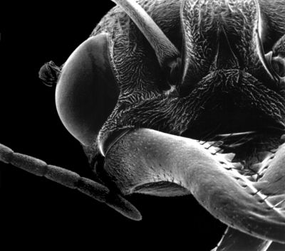
Open mouthparts of bark-inhabiting ant species Melissotarsus sp. from Africa. See on Sketchfab. See list of 3D images.