Key to Subfamilies of North America
This key to the subfamilies of North America has been adapted from Fisher & Cover (2007).
1
- Upper plate (tergite) of the last abdominal segment (pygidium) flattened and with a pair of distally converging rows of spines or peg-like teeth (A) => Dorylinae (Genus key)
- Pygidium rounded and without a pair of distally converging rows of spines or peg-like teeth => 2
2
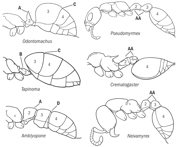
- Postpetiole absent; waist consisting of a petiole (abdominal segment 2) (A). In some cases, petiole greatly reduced and node vestigial or absent (Tapinoma, Technomyrmex) (B). In profile, abdominal segment 3 not markedly smaller in size than abdominal segment 4 and either confluent with (C) or separated from it by a slight or moderate constriction (D) => 3
- Postpetiole present; waist consisting of two segments (AA). Following the petiole is the postpetiole (abdominal segment 3), which is much smaller than abdominal segment 4 and separated from it by a strong constriction => 5
3
- Sting absent. No constriction present between abdominal segments 3 and 4 (A) => 4
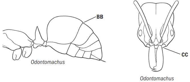
- Sting present, often prominent. Usually with a visible constriction between abdominal segments 3 and 4 (AA); if without a visible constriction (Anochetus, Odontomachus) (BB), then mandible inserted in the middle of the front margin of the head (CC) => 7
4
- Apex of abdomen with a circular opening (acidopore), usually surrounded by a fringe of hairs (A). If the fringe of hairs is not present, then the antennal insertions are set well back from the posterior border of the clypeus (Camponotus) (B) => Formicinae (Genus key)
- Acidopore absent. Apex of abdomen with a transverse slit-like orifice, not surrounded by a fringe of hairs (AA). Antennal insertions set at or directly adjacent to the posterior border of the clypeus (BB) => Dolichoderinae (Genus key)
5
- Combining the following characters: First mesosomal segment freely articulating with the second mesosomal segment, forming a flexible joint (A). Tibia of hind leg with a prominent, pectinate spur (use high magnification, 50 to 100 x) (B). Eyes unusually large, often covering most of the sides of the head (C). Ocelli present => Pseudomyrmecinae (Pseudomyrmex, Species key)
- First mesosomal segment fused with the second mesosomal segment, forming an inflexible structure (AA). Hind tibial spurs usually simple (BB), sometimes pectinate (BBB) or absent. Eyes variously developed, sometimes vestigial or absent. When present, seldom unusually large, never covering most of the sides of the head. Ocelli nearly always absent => 6
6
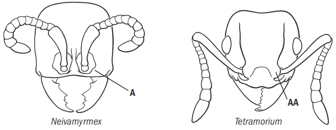
- Antennal sockets completely exposed in full-face view, not covered by frontal lobes (A); antennal sockets closely approximated, inserted at the anterior margin of the head (A). Clypeus reduced. Eyes either a single facet or absent => Dorylinae (Genus key)
- In full-face view, antennal sockets covered at least in part by frontal lobes (AA); antennal sockets usually well separated and set back from the anterior margin of the head at or near the posterior border of the clypeus (AA). Clypeus developed. Eyes usually present, often with several facets or more (BB) => Myrmicinae (Genus key)
7
- Anterior margin of clypeus denticulate (A). Petiole broadly attached to third abdominal segment (B); petiole with distinct front and dorsal faces but without a distinct sloping posterior face; petiole separated at most by a shallow impression from third abdominal segment => Amblyoponinae (Genus key)
- Anterior margin of clypeus not denticulate (AA). Petiole narrowly attached to third abdominal segment (BB); petiole with distinct front, top, and posterior faces; petiole’s attachment to the third abdominal segment narrow and strongly constricted => 8
8
- Upper plate (tergite) of abdominal segment 4 coarsely and longitudinally costate (A). Hind coxa with dorsal spine (B). Tarsal claws always with a single subapical tooth, in addition to the terminal point (C) => Ectatomminae (Gnamptogenys, Species key)
- Tergite of abdominal segment 4 lacking sculpture or with fine textured sculpture, never with striate or costate sculpture (AA). Hind coxa lacking dorsal tooth or spine (BB). Tarsal claws usually simple (CC) => 9
9
- Dorsum of mesosoma lacking a promesonotal suture, with the metanotal suture usually lacking as well (A) (metanotal groove present in Proceratium californicum). Tergite of abdominal segment 4 always enlarged and strongly arched so that it forms the rearmost part of the abdomen when viewed in profile; apex of abdomen directed anteriorly (B) => Proceratiinae (Genus key)
- Dorsum of mesosoma with at least the promesonotal suture; the metanotal suture usually present as well (AA). Abdominal tergite 4 not strongly enlarged and only weakly arched; apex of abdomen directed rearwards or ventrally, not anteriorly (BB) => Ponerinae (Genus key: Fisher and Cover, Schmidt and Shattuck)
References
- Fisher, B. L.; Cover, S. P. 2007. Ants of North America. A guide to genera. Berkeley: University of California Press, xiv + 194 pp.


