Martialis heureka
| Martialis heureka | |
|---|---|

| |
| Scientific classification | |
| Kingdom: | Animalia |
| Phylum: | Arthropoda |
| Class: | Insecta |
| Order: | Hymenoptera |
| Family: | Formicidae |
| Subfamily: | Martialinae |
| Genus: | Martialis |
| Species: | M. heureka |
| Binomial name | |
| Martialis heureka Rabeling & Verhaagh, 2008
| |


On the basis of the specimen’s external morphology, we are able to infer some aspects of the species’ biology. The pale integument and the absence of eyes suggest that M. heureka lives hypogaeically or in covered low-light environments, like leaf litter or rotting wood. The fact that the first two M. heureka individuals were collected in soil core samples during the day, and the present specimen in leaf litter at dusk, supports this hypothesis. Possibly, M. heureka surfaces during the night to forage. The unusually enlarged procoxae and long front legs could potentially be an adaptation to prey capture. Presumably, they are used less for digging activities, because the legs are relatively thin and lack the characteristic erect setae of actively digging species. We speculate that M. heureka might take advantage of preexisting underground cavities, like hollow rotten roots. The forceps-like mandible type is not seen in any other ant species. These long, filigree instruments could be used to drag soft prey items out of cavities. Annelids, termites, insect larvae, and other soft-bodied arthropods are possible prey. We do not expect M. heureka to prey on heavily sclerotized invertebrates. (Rabeling et al. 2008)
Identification
See the description given below.
Distribution
Latitudinal Distribution Pattern
Latitudinal Range: -2.37204° to -2.883333333°.
| North Temperate |
North Subtropical |
Tropical | South Subtropical |
South Temperate |
- Source: AntMaps
Distribution based on Regional Taxon Lists
Neotropical Region: Brazil (type locality).
Distribution based on AntMaps
Distribution based on AntWeb specimens
Check data from AntWeb
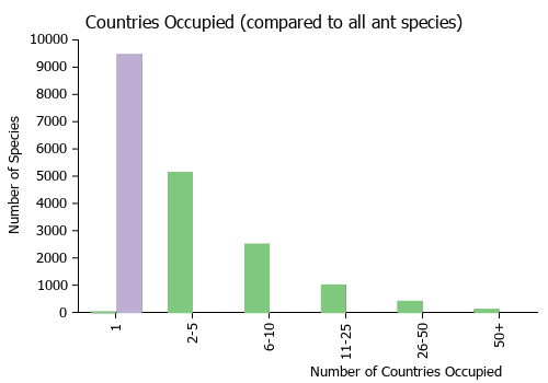
Countries Occupied
| Number of countries occupied by this species based on AntWiki Regional Taxon Lists. In general, fewer countries occupied indicates a narrower range, while more countries indicates a more widespread species. |
|
Estimated Abundance
| Relative abundance based on number of AntMaps records per species (this species within the purple bar). Fewer records (to the left) indicates a less abundant/encountered species while more records (to the right) indicates more abundant/encountered species. |

|
Biology
Boudinot (2015)
This species was described from a single stray worker from the Amazon just north of Manaus, Brazil, Martialis heureka is one of the most significant taxa in the Formicidae described in recent years. Displaying a bizarre mixture of pleisiomorphic and autapomorphic traits, the species was attributed to its own subfamily, the Martialinae. This decision was supported by further morphological study (Brandão et al. 2010) and multi-locus molecular phylogenetic reconstruction (Rabeling et al. 2008; although see Kück et al. 2011). Rabeling et al. (2008) recovered Martialis as the sister to all remaining extant ants including the Old World subfamily Leptanillinae, while a reanalysis by Kück et al. (2011) found the converse. Given this debate and the mysterious biology of the species, Martialis is of high interest. Here the male of Martialis is described for the first time based on material from the Biological Dynamics of Forest Fragments Project (BDFFP [English], PDBFF [Portuguese]) study region, about 50 km north of the type locality.

The male of Martialis differs from the worker by standard intercaste dimorphism, e.g., eyes well-developed, ocelli and notauli present, alate, flight sclerites developed, mesosoma musculated for flight, but the male also differs notably in several specific characters: mandibles far shorter, reduced relative to worker; labral trigger setae absent; clypeal brush absent; antennal toruli situated more anteriorly; forelegs weak; metatibial gland absent; petiolar node weakly developed; helcium broader; abdominal tergum and sternum III of equal length; cinctus between pre- and postsclerites IV not developed. On the other hand, the male of Martialis displays numerous similarities with the worker, such as linear mandibles, reduced palpal count (although worker count unconfirmed, but certainly less than 3,3), clypeus poorly developed, anterior tentorial pits posteriorly-situated, antennal toruli well-developed (but not quite as cup-like as in worker), scapes long, pedicel elongate, frontal carinae absent, propodeal lobes weakly developed (contra the initial worker diagnosis), tibial spur formula 1,1, basipetiolar carina present, petiolar tergum and sternum fused, abdominal segment III differentiated from IV, and sculpture and setation remarkably similar, although the somatic sclerites of the male are generally less strongly sclerotized. The two castes are similar in several other specifics, but this brief list captures most of the significant features.
Specimens were examined from collecting events in January, February, April, and October of 1985. The BDFFP study region displays weak seasonality of rainfall and day length, with the months of June through December roughly representing the “dry season”, July through September being the driest (Bierregaard Jr. et al. 2001). It is possible that Martialis flights occur year-round, although the sampling of BDFFP material examined for this study is too small to confidently assert the flight phenology. While the range of Martialis has only been extended by about 50 km by the discovery of the male, the quantity of males recovered exceeds that of workers by an order of magnitude (25 males vs. 3 workers). Thus, Martialis may be recovered via alates more readily than workers. The use of Malaise traps and flight intercept traps should be encouraged for studies of ant diversity, particularly for species with cryptic habits. Although our knowledge of flight phenology is poor, tropical rainforests may be particularly amenable to these studies due to the relatively more year-round flights of Neotropical (Kaspari et al. 2001a, 2001b) than of Nearctic ants (Dunn et al. 2007).

As the Manaus region is considered the ecological “crossroads” of the Amazon where a high proportion of species ranges overlap (Bierregaard Jr. et al. 2001), it will be valuable to sample for Martialis in other Amazonian regions. Moreover, it is of interest whether the fragmented populations of Martialis in the BDFFP plots have survived the intervening 30 years. This may not be the case, as ant communities have been observed to hemorrhage in BDFFP study plots (Vasconcelos et al. 2001), although hypogaeic ants may be less sensitive to habitat changes than epigaeic ants. The placement of the Malaise samples relative to the edges of the study plots is unknown. As well, the gyne of Martialis remains unknown. It is possible that this caste will be ergatoid, but the presence of alate gynes cannot be ruled out. In general, the male of a given ant species is more frequently collected via Malaise traps than females; thus it is possible that although only males were encountered, alate gynes could still be present. Regardless, the natural history of Martialis will be fascinating to uncover.
Castes
Known from workers and males (images below in description).
Nomenclature
The following information is derived from Barry Bolton's Online Catalogue of the Ants of the World.
- heureka. Martialis heureka Rabeling & Verhaagh, in Rabeling, et al. 2008: 14914, figs. 1, 2 (w.) BRAZIL (Amazonas).
- Type-material: holotype worker.
- Type-locality: Brazil: Amazonas, Manaus, Headquarters of Empresa Brasileira de Pesquisa Agropecuária (EMBRAPA), Amazônia Occidental, at km. 28 of highway AM010, 2°53’S, 59°59’W, 40-50 m., 9.v.2003, primary tropical lowland rainforest, ex leaf litter (C. Rabeling).
- Type-depository: MZSP.
- Boudinot, 2015: 37 (m.).
- Status as species: Brandão, et al. 2010: 413; Kück, et al. 2011: 1; Boudinot, 2015: 37.
- Distribution: Brazil.
Unless otherwise noted the text for the remainder of this section is reported from the publication that includes the original description.
We assume that the present specimen of M. heureka is a worker, because it lacks ocelli, the enlarged mesosoma and the extra sclerites associated with wings. In addition, the specimen was collected in the leaf litter suggesting foraging activity usually performed by the worker caste. However, queens with worker-like morphology have been reported from several poneroids and socially parasitic Formicinae and Myrmicinae. Because we did not perform a dissection of the single specimen no statement can be made about palpal segmentation or internal anatomy.
Description
Worker
(holotype). Measurements:HW0.65 mm, HL 0.62 mm, SL 0.46 mm, FL 1.03 mm, ML 0.90 mm, WL 1.02 mm, PW 0.40 mm, PEW 0.19 mm, PEL 0.27 mm, PPW 0.34 mm, PPL 0.30 mm, HFL 0.60 mm, HTL 0.57 mm, CI 105, MI 145, SI 72, DI 45. Including the characters of the subfamily and genus description: small (HW 0.65, WL 1.02), pale yellow, and blind, integument in dried condition partly translucent. Very long, slender, forcepslike mandibles (ML 0.90, MI 145) inserted on outer anterior margin of head capsule, projecting straight forward to ~5/7 of its length, then curving slightly mesally. Inner margin bearing a tiny sudmedian tooth at ~2/5 of its length, measured from the base and a group of 3 subapical teeth at ~5/7 of its length: a median larger tooth and a somewhat smaller tooth to each side of it. The subapical teeth and the sharply pointed mandibular tip frame an oval space. Mandibles crossed in dried condition, not crossing in live individual or while stored in 80% EtOH. Head capsule bulbous in lateral view, narrowing toward posterior margin. Clypeus reduced, narrow strip between lateral head margin and wider between antennal sockets, covered with at least 20 setae that project forward, beyond the margin of antennal sockets, resembling a brush. Antennal scape relatively short (SI 72, DI 45), 1st and 2nd funicular segment 3⁄4 and 1⁄4 longer than 3rd funicular segment. Funiculus more than twice as long as scape. Mesosoma long and slender. Promesonotal suture present, pronotum and mesonotum presumably capable of movement relative to each other. Pronotum forming a slender cervix. Front legs enlarged: procoxae twice as long and wide as meso and metacoxae; profemura and tibiae also enlarged. Petiole subsessile with a short anterior peduncle; petiolar node rounded, smooth, with a short sloping posterior face; no teeth or projection ventrally. Metasoma (abdominal segment IV–VIII visible) laterally compressed, drop shaped in lateral view. Head, pronotum, and legs densely covered with erect to suberect hairs and sparsely with longer erect setae; mandibular pubescence dense, consisting of short suberect hairs. Inner margin with two rows of at least 18 long straight setae, which are arranged pair wise. Propodeum without any hairs; petiole with few suberect setae on dorsal surface of node and abdominal segment III–VIII with irregularly spaced long erect setae. Dense appressed pubescence absent from entire body. Only few body parts bear distinct sculpturing: neck, mesonotum, propodeum, and ventral surface of petiole punctate; lateral surface of propodeum faintly striate.
Male

Boudinot (2015) - (n=3). HL 0.35–0.42, HW1 0.35–0.40, HW2 0.44–0.49, MAL 0.04–0.06, MDL 0.10–0.11, SL 0.20–0.23, PDL 0.10–0.13, A3L 0.17–0.21, AAL 0.17–0.21, EL 0.17–0.19, EW 0.14–0.17, OOD 0.14–0.15, LOD 0.04–0.05, MOD 0.04–0.05, ML 0.64–0.78, MLL 0.16–0.18, MLW 0.17–0.20, MTL 0.29–0.37, MTW 0.36–0.45, PFL 0.39–0.48, MFL 0.43–0.53, PTH 0.16–0.19, PTL 0.20–0.23.
Indices. CI 0.94–1.02, CS 0.35–0.41, SEI 83.6–86.2, SI 55.6–58.4, EI 83.0–85.6, EYE 88.1–88.7, MI 26.7–27.8, OBI 80.1–82.8, OMI 3.46–4.74, MNI 2.00–2.26, MTI 79.9–83.2, FI 88.3–91.3, PTI 76.9–83.2. Small, but body variable in overall size (Fig. 11C).
Head (Fig. 11A–B). In full-face view head about as broad as long excluding eyes, broader than long including eyes. Palpal formula 2,1; palps short, not reaching hypostomal margin. Stipes simple, lacking carinae on medial surface. Labrum very small, medially emarginate, setose; lateral margins distant from mandibular bases by somewhat less than maximum lateromedial labrum length; labrum lacking basolateral trigger setae observable in workers. Mandibles linear, narrow; lateral and medial margins weakly tapering to apex; masticatory mandibular margin reduced, bidentate; apical tooth asymmetrical, larger than symmetrical basal tooth; mandalus enlarged, diameter equal to maximum mandible width. Clypeus reduced; anterior margin broadly emarginate; medial clypeal portion maximum anteroposterior length about 1.5 maximum antennal socket diameters; posterior clypeal margin produced between antennal toruli. Supraclypeal area arc-shaped, anteroposteriorly longer than maximum antennal socket diameter. Antennal toruli situated anteriorly, with anteriormost portion of torular arch anterad anterior tentorial pit. Frons and ocellar area bulging. Occipital carina present, weakly developed, obscured in full-face view by vertex, not enclosing occiput. Compound eyes bulging strongly; medial margin weakly convex; posterior margin weakly emarginate; compound eye narrower dorsally than ventrally. Ocelli small, situated distant from compound eye. Hypostomal margin reduced, lacking lamina. Antenna 13-merous; scape longer than maximum compound eye diameter and slightly more than 2 x pedicel length; pedicel cylindrical, long, about 4/5 x antennomere 3 length; funiculus filiform, elongate, reaching metasoma when laid against mesosoma.
Mesosoma (Fig. 11C–D). Pronotal neck continuous with remainder of sclerite in dorsal view; main portion of pronotum swollen, muscular; anteromedian pronotal face convex in profile view, short, dorsoventral height of pronotum from pronotal neck about 1/3 x mesoscutum height in profile view; lateral pronotal face concave. Mesoscutum broader than long in dorsal view (length 0.80–0.83 x width); anterior and posterolateral areas swollen. Notauli distinct, crossribbed, meeting at body midline, not extending to transscutal line although narrow longitudinal line present from notauli to transscutal line. Parapsidal lines impressed, slightly divergent. Parascutal carinae nearly linear; weakly sinuate. Scutoscutellar sulcus unimpressed. Axillae small and widely situated. Mesoscutellum high and convex in profile view; not modified. Metascutellum small, lateromedial width slightly less than one half anteroposterior length; in profile view metascutellum strongly produced. Metanotal trough deep, small, circular. Mesopectus with oblique longitudinal sulcus, anterior terminus of sulcus nearly contacting posterolateral pronotal corner. Spiracular sclerite inconspicuous. Lower metapleural area strongly offset from upper metapleural area by deep, broad, margined sulcus. Metapleural gland orifice occluded; presence of internal metapleural gland not visible through metapleural sclerite. Propodeum parabolic in profile view, dorsal face about as long as and continuous with posterior face; propodeal spiracle circular, small; propodeal lobe weakly developed, carinate, clearly visible in anterolateral oblique view.
Metasoma (Fig. 11C). Petiole nodiform, pedunculate; anteriormost portion of petiolar tergum offset by parabolic carina; petiolar tergum and sternum fused, longitudinal lateral carinae not suggestive of suture; petiolar node shallow, in profile view anterodorsal face nearly linear, dorsum weakly convex, posterior face very weak; petiolar sternum linear for most of length, posteriorly narrowed, ventral petiolar surface with paired diverging carinulae; subpetiolar process absent. Abdominal segment III slightly reduced and differentiated from segment IV; helcium axial, sternal presclerite visible in profile view, not obscured by tergal presclerite; abdominal posttergite and poststernite III not fused; abdominal sternum III prora present as anterolateral bosses subtending helcium, anteromedian area of sternum concave. Abdominal terga IV–VIII and abdominal sterna IV–IX normally developed, not reduced or obscured in situ. Abdominal tergum VIII posterior margin unmodified. Abdominal sternum IX apically ligulate, narrow, posterior margin very narrowly convex, nearly triangular.

Forewing (Fig. 12A). Tegulum reduced, subrectangular, longer than broad. Wings weakly infuscated, completely covered in fine setose layer. Pterostigma poorly-developed, only anterior enclosing abscissa tubular. Wing venation Ogata type IVa: Submarginal cell 1+2 and marginal cell 1 closed, 1m-cu absent, thus discal cell 1 open. Costal vein tubular to pterostigma. Rsf1 slightly more than 1/2 x length of and meeting Mf1 obliquely. Rs+M continuous with undifferentiated Mf2-3 until meeting very short 2rsm. Rsf2+3 absent. 2r-rs very long, longer than combined lengths of Rsf1 and Mf1; 2r-rs directed posteroapically, not orthogonal with anterior wing margin. Rsf4–6 tubular to Rf, enclosing marginal cell. Mf4–6 absent. Crossvein cu-a incompletely tubular, situated basad Mf1. Cuf divergent with respect to Rs+M+Mf2+3. 1A extending only slightly beyond cu-a, not enclosing subdiscal cell 1.
Hindwing (Fig. 12B). Hindwing venation reduced, only R+Rs and 1A tubular; R not reaching anterior wing margin; 1A short, weakly indicated. Three hamuli present. Claval region poorly developed.
Genitalia (Fig. 12C–H). Pygostyles absent. Abdominal sternum IX spiculum short; anterior margin linear, curving posterolaterally near lateral margins; lateral margins short, slightly divergent; posterolateral margins weakly concave, tapering strongly to acute, narrowly rounded apex. Cupula dorsal and lateral faces about as broad as telomeral base; lateral face narrowing ventrally to narrow bar-like ventral face. Basimere and telomere more-or-less continuous, basimere weakly shouldered dorsomedially anterad telomeral base; dorsomedian margins of basimeres parallel for about half length of paramere; telomere acutely triangular in profile view; basimere and telomere with ventrolateral layer of posteroventrallydirected setae. Basivolsella lateromedially broad; base transversely connected with basimere; cuspis absent; digitus clavate, apex swollen and directed ventrally; digital stem short. Valvura short, linear, directed anteriorly and situated at about 2/3 valviceps height; valviceps linear, dorsoventrally short; valviceps dorsomedially fused for most of length; in profile view dorsal valviceps margin weakly convex, ventral margin concave, edentate, valviceps apex weakly convex, subrectangular, produced ventrally; dorsolateral valviceps face convex, margined by lateral apodeme which extends almost to apex before curving ventrad and contacting ventral margin; ventrolateral valviceps face concave; phallotreme situated at aedeagal apex, sclerotic aperture formed by valviceps circular.
Coloration. Body almost uniformly brown to brownish yellow; extremities slightly lighter colored.
Sculpturation. Body weakly sculptured overall; head with fine piligerous punctae; mesonotal piligerous punctae coarser, posterolateral mesoscutal area above parascutal carina roughened; dorsomedian scutoscutellar area finely and densely anteroposteriorly striate; mesoscutellum weakly roughened; metascutellum with fine transverse carina subtending posteriorly produced portion of disc; mesopectus and metapleuron smooth, shining, slightly rough; propodeum finely striate, striae extending from anterior margin down along lateral faces, dorsal propodeal face weakly rugose, posterior face mostly smooth; petiole mostly smooth and shining, with lateral longitudinal carinulae; abdominal segment III mostly smooth and shining; abdominal segments posterior to segment III weakly sclerotized.
Setation. Head, median pronotal portion including pronotal neck, mesonotum, and procoxae covered by dense layer of somewhat short, uniform, weakly curved, erect to suberect setae, longer setae present on these areas very sparsely; clypeus lacking clypeal brush of worker, although setal layer denser than on remainder of head capsule; setae sparse and subdecumbent to nearly appressed on pronotal lateral face, mesopectus, metapleuron, and propodeum; setae somewhat denser on metasoma, but not as dense as on head and mesonotal dorsum; petiolar setae elongate, linear, setae on remaining segments shorter and curved; setae on legs, including meso- and metacoxae, about as dense as on metasoma, mostly subdecumbent with a few longer suberect setae present.
Type Material
Brazil: Amazonas, Manaus. Headquarters of Empresa Brasileira de Pesquisa Agropecua´ria (EMBRAPA)-Amazonia Ocidental, located at kilometer 28 of highway AM 010; 2°53'S, 59°59'W; elev. 40–50 m; 09 May 2003; col. C. Rabeling; ex leaf litter at dusk, primary tropical lowland rainforest. The holotype is deposited in Museu de Zoologia daUniversidade de Sa˜o Paulo, Brazil (MZSP).
Etymology
The species epithet heureka (gr.:I found it!) epitomizes the troubled story of the species’ rediscovery. Five years after two workers were discovered by M. Verhaagh in a soil sample and subsequently lost, a single worker, the present holotype, was recollected in a nearby patch of primary rainforest.
References
- Borowiec, M.L., Moreau, C.S., Rabeling, C. 2020. Ants: Phylogeny and Classification. In: C. Starr (ed.), Encyclopedia of Social Insects (doi:10.1007/978-3-319-90306-4_155-1).
- Boudinot, B.E. 2015. Contributions to the knowledge of Formicidae (Hymenoptera, Aculeata): a new diagnosis of the family, the first global male-based key to subfamilies, and a treatment of early branching lineages. European Journal of Taxonomy 120, 1-62 (http://dx.doi.org/10.5852/ejt.2015.120).
- Brandão, C.R.F., Diniz, J.L.M. & Feitosa, R.S.M. 2010. The venom apparatus and other morphological characters of the ant Martialis heureka. Papeis Avulsos de Zoologia (São Paulo) 50:413-423.
- Fernandez, F., Guerrero, R.J., Sánchez-Restrepo, A.F. 2021. Sistemática y diversidad de las hormigas neotropicales. Revista Colombiana de Entomología 47, 1–20 (doi:10.25100/socolen.v47i1.11082).
- Griebenow, Z. 2020. Delimitation of tribes in the subfamily Leptanillinae (Hymenoptera: Formicidae), with a description of the male of Protanilla lini Terayama, 2009. Myrmecological News 30: 229-250. (doi:10.25849/MYRMECOL.NEWS_030:229).
- Griebenow, Z.H. 2021. Synonymisation of the male-based ant genus Phaulomyrma (Hymenoptera:Formicidae) with Leptanilla based upon Bayesian total-evidence phylogenetic inference. Invertebrate Systematics 35, 603–636 (doi:10.1071/is20059).
- Griebenow, Z.H., Isaia, M., Moradmand, M. 2022. A remarkable troglomorphic ant, Yavnella laventa sp. nov. (Hymenoptera: Formicidae: Leptanillinae), identified as the first known worker of Yavnella Kugler by phylogenomic inference. Invertebrate Systematics 36(12), 1118-1138 (doi:10.1071/is22035).
- Houadria, M., Menzel, F. 2021. Digging Deeper into the Ecology of Subterranean Ants: Diversity and Niche Partitioning across Two Continents. Diversity 13, 53 (doi:10.3390/d13020053).
- Jansen, G., Savolainen, R. 2010. Molecular phylogeny of the ant tribe Myrmicini (Hymenoptera: Formicidae). Zoological Journal of the Linnean Society 160(3), 482–495 (doi:10.1111/j.1096-3642.2009.00604.x).
- Kück, P., Hita Garcia, F., Misof, B. & Meusemann, K. 2011. Improved phylogenetic analyses corroborate a plausible position of Martialia heureka in the Ant Tree of Life. PLoS ONE 6 (6): 1-8 (e21031. doi: 10.1371/journal.pone.0021031).
- Perfilieva, K.S. 2023. Cretaceous-Burmese-amber ants: Morphological features and community structure. Biology Bulletin Reviews 131, 38–54 (doi:10.1134/s207908642301005x).
- Rabeling C, Brown JM and Verhaagh M. 2008. Newly discovered sister lineage sheds light on early ant evolution. Proceedings of the National Academy of Sciences. 105(39):14913-14917.
- Richter, A., Boudinot, B., Yamamoto, S., Katzke, J., Beutel, R. G. 2022. The first reconstruction of the head anatomy of a Cretaceous insect, †Gerontoformica gracilis (Hymenoptera: Formicidae), and the early evolution of ants. Insect Systematics and Diversity 6(5): 1-80 (doi:10.1093/isd/ixac013).
- Romiguier, J., Borowiec, M. L., Weyna, A., Helleu, Q., Loire, E., La Mendola, C., Rabeling, C., Fisher, B. L., Ward, P. S., Keller, L. 2022. Ant phylogenomics reveals a natural selection hotspot preceding the origin of complex eusociality. Current Biology, 3213, 2942-2947.e4 (doi:10.1016/j.cub.2022.05.001).