Key to Subfamily of Philippine Ants
Based on the worker caste (adapted from Bolton, 1994).
Return to Philippine Ants or List of ant species of the Philippines.
1
- Postpetiole absent (Fig. 1a) . . . . . 2
- Postpetiole present and segments nearly equal in size, or postpetiole much larger, in which case, gaster with distinct constriction at insertion with postpetiole (Fig. 1b) . . . . . 7
 1a. Profile of Anoplolepis gracilipes worker |
 1b. Profile of Calyptomyrmex loweryi worker |
2
( 1 )
- Sting absent . . . . . 3
- Sting present, often conspicuous and functional . . . . . 4
3
( 2 )
- In profile, a circular nozzle, the acidopore, often fringed with hairs, present at apex of gaster; antennal sockets sometimes situated well behind posterior margin of clypeus Formicinae
- In profile, acidopore absent; apex of gaster with a transverse slit-like orifice (best seen in apical or lateral view); antennal sockets always abutting the posterior margin of clypeus Dolichoderinae
4
( 2 )
- Petiole broadly attached to gaster, so that petiole has no posterior face in side view (Fig.4a); mandibles linear and inserted at side of head (Fig. 5a) Amblyoponinae
- Petiole narrowly attached to gaster, so that petiole has a posterior face in side view (Fig. 4b); mandibles variable and inserted at sides or front of head (Fig. 5b) . . . . . 5
 4a. Profile of Stigmatomma rothneyi worker |
 4b. Profile of Ooceraea biroi worker |
 5a. Frontal view of Stigmatomma rothneyi worker |
 5b. Frontal view of Ooceraea biroi worker |
5
( 4 )
- Frontal lobes and clypeus absent or reduced, so that antennal sockets are fully exposed in dorsal view and situated very near or above anterior margin of the head (Fig. 6a) Proceratiinae
- Frontal lobes and clypeus well-developed, so that antennal sockets are partially or entirely covered in dorsal view and situated posterior to the anterior margin of the head (Fig. 6b) . . . . . 6
 6a. Frontal view of Proceratium papuanum worker |
 6b. Frontal view of Stictoponera menadensis worker |
6
( 5 )
- Frontal lobes elongate and roughly parallel, so that frontal carinae do not converge posteriorly; mandibles triangular (Fig. 7a) Ectatomminae
- Frontal lobes rounded or bluntly triangular; frontal carinae, when present, appear to converge posteriorly; mandibles variable from linear to triangular (Fig. 7b) Ponerinae
 7a. Profile of Rhytidoponera araneoides worker |
 7b. Profile of Myopias breviloba worker |
7
( 1 )
- Pronotum and mesonotum separated by a conspicuous, flexible suture, allowing pronotum to move relative to mesonotum (Fig. 8a) . . . . . 8
- Pronotum and mesonotum separated by an often inconspicuous, rigid suture, so that pronotum and mesonotum are fused together (Fig. 8b) . . . . . 9
 8a. Profile of Tetraponera nitida worker |
 8b. Profile of Paratopula macta worker |
8
( 7 )
- Eyes, present, large; ventral surface of postpetiole at most slightly bulging (Tetraponera) (Fig. 9a) Pseudomyrmecinae
- Eyes absent; ventral surface of postpetiole swollen and bulbous (Leptanilla) (Fig. 9b) Leptanillinae
 9a. Profile of Tetraponera nigra worker |
 9b. Profile of Leptanilla revelierii worker |
9
( 7 )
- Postpetiole barrel-shaped and much larger than petiole; gaster conspicuously constricted at insertion; tergite VII transversely flattened and armed with peg-like teeth (Fig. 10a) Cerapachyinae
- Postpetiole globular and nearly equal in size to petiole; gaster with narrow constriction at insertion; tergite VII rounded and unarmed (Fig. 10b) . . . . . 10
 10a. Profile of Cerapachys jacobsoni worker |
 10b. Profile of Eurhopalothrix procera worker |
10
( 9 )
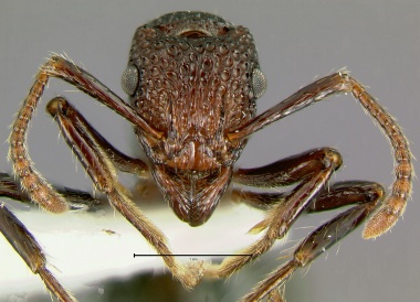
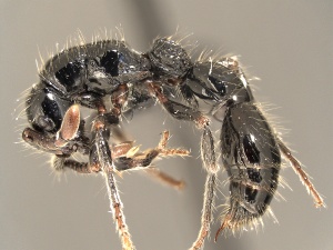
- Frontal lobes absent; antennal sockets completely exposed; eyes absent (Fig. 11a); gaster narrowly constricted at insertion with postpetiole (Aenictus) (Fig. 12a) Aenictinae
- Frontal lobes usually present (much reduced or absent in Pristomyrmex)(Fig. 11b); antennal sockets partially or completely concealed; eyes rarely absent; gaster not constricted at insertion with postpetiole (Fig. 12b) Myrmicinae
 11a. Frontal view of Aenictus nesiotis worker |
 11b. Frontal view of Pristomyrmex quadridens worker |
 12a. Profile of Aenictus nesiotis worker |
 12b. Profile of Pristomyrmex quadridens worker |