Zasphinctus lumumbai
| Zasphinctus lumumbai | |
|---|---|

| |
| Scientific classification | |
| Kingdom: | Animalia |
| Phylum: | Arthropoda |
| Class: | Insecta |
| Order: | Hymenoptera |
| Family: | Formicidae |
| Subfamily: | Dorylinae |
| Genus: | Zasphinctus |
| Species group: | obamai |
| Species: | Z. lumumbai |
| Binomial name | |
| Zasphinctus lumumbai Hita Garcia & Gómez, 2025
| |


Zasphinctus lumumbai is so far only known from one specimen in the MRAC collection, so our knowledge is limited to it being found at the type locality in forest soil.
Photo Gallery
 Hita Garcia et al. (2025), Figure 22. Shaded surface display volume renderings of 3D models of Z. lumumbai holotype (MRACFOR0010007). A, full body in profile. B, full body in dorsal view. C, head in full-face view (with antennae). D, head in full-face view (without antennae). E, head in ventral view. F, abdominal segment II (petiole) in profile. G, abdominal segment II (petiole) in dorsal view. H, tergum of AS III in dorsal view I sternum of AS III in ventral view.
,_Fig._22.jpg)
Identification
With characters of the Z. obamai group (Figs 19D, F, H, 20D, F, H):
- body size significantly smaller (HL 0.54–0.60; WL 0.73–0.87)
- head in full-face view appearing thinner (CI 78–80)
- head in profile appearing conspicuously thinner, its underside only slightly curved
- clypeal area always without conspicuous median tooth
- torular-posttorular complex in dorsal view with sides more or less parallel
- torular-posttorular complex in profile strongly arched anteriorly towards highest dorsal point and posterodorsally lobate
- vertexal margin in posterodorsal view strongly developed delimiting posterior face of head
- anterior outline of occipital margin in ventral view moderately or weakly and irregularly defined and with anterolateral projections angulate (in Z. obamai rounded)
- vertex with clear margin laterally and not appearing fused to the occiput
- anterior outline of postoccipital margin in ventral view moderately or weakly and irregularly defined and with anterolateral projections angulate; mesosoma in profile relatively lower and elongate (LMI 34–37)
- pleural endophragmal pit strongly developed and deep
- petiolar tergum in profile relatively lower (LPI 112-123)
- mesosoma in dorsal view appearing thinner and elongate (DMI 38–40; DMI2 49–53)
- petiolar tergum in profile relatively lower, ~ 0.8–0.9 × higher than long (LPI 112–123)
- petiolar tergum in dorsal view thinner, ~ 0.8–0.9 × broader than long (DPI 82–93)
- abdominal tergum III in dorsal view strongly trapezoidal with anterior margin more angulate
- abdominal sternum III in ventral view rounded trapezoidal, comparatively thinner and higher, sides less rounded
- usually with conspicuous surface sculpture somewhere on body (except for piliferous foveae), usually on cephalic dorsum and sides of mesosoma
Plus the following:
- body size significantly much smaller (HL 0.54; WL 0.73)
- lateral arms of hypostomal carina less diverging, relatively thin, and angulate at widest points (Fig. 8D)
- postgenal sulcus restricted to area adjacent to hypostomal carina and only weakly impressed (Fig. 8D)
- postoccipital margin in ventral view with anterior outline moderately or weakly and irregularly defined
- anterolateral projections angulate (Fig. 8D)
- pleural endophragmal pit weakly developed and shallow but visible (Fig. 10D)
- subpetiolar process of petiole (AS II) in profile with thickened anterior and ventral margins and weak concavity without differentiated fenestra (Fig. 13D)
- posterior end of abdominal segment III in ventral view with thick, deep, sharply and irregularly outlined transverse groove (Fig. 16D)
- prora in anteroventral view well-developed with thick, irregularly shaped and rounded lateroventral margins (Fig. 16D)
- surface sculpture on cephalic dorsum and genae mostly smooth and shiny with abundant, relatively deep, and large piliferous foveae, except for reticulate–punctate anteromedian area (Figs 4D, 5D, 19D, 20D)
- general surface sculpture on mesosoma and metasoma seemingly smooth and shiny with varying degrees of scattered piliferous foveae, hypopygidium reticulate-rugose (Figs 20D, 21D).
[General surface sculpture difficult to assess since larger areas are covered in glue and dirt.]
 Hita Garcia et al. (2025), Figure 4. Diagnostic plate showing still images from surface volume renderings of the head in full-face view (the remainder of the body virtually removed) (torular-posttorular complex in semi-transparent red; anterior projections of parafrontal ridges in semi-transparent yellow). A, Z. aprilia holotype (CASENT0764763). B, Z. kouakoui paratype (CASENT0764653). C, Z. lolae holotype (KGCOL02270). D, Z. lumumbai holotype (MRACFOR0010007). E, Z. ndouri holotype (KGCOL01883). F, Z. obamai holotype (CASENT0764125). G, Z. sarowiwai holotype (CASENT0764654). H, Z. wilsoni holotype (MCZ-ENT00512764).
 Hita Garcia et al. (2025), Figure 5. Diagnostic plate showing still images from surface volume renderings of the head in profile view (the remainder of the body virtually removed) (torular-posttorular complex in semi-transparent red). A, Z. aprilia holotype (CASENT0764763). B, Z. kouakoui paratype (CASENT0764653). C, Z. lolae paratype (CASENT0764651). D, Z. lumumbai holotype (KGCOL02270). E, Z. ndouri holotype (KGCOL01883). F, Z. obamai holotype (CASENT0764125). G, Z. sarowiwai holotype (CASENT0764654). H, Z. wilsoni holotype (MCZ-ENT00512764).
 Hita Garcia et al. (2025), Figure 6. Diagnostic plate showing still images from surface volume renderings of the head in posterodorsal view (the remainder of the body virtually removed) (occipital margin in semi-transparent red; vertexal margin in semi-transparent yellow). A, Z. aprilia holotype (CASENT0764763). B, Z. kouakoui paratype (CASENT0764653). C, Z. lolae holotype (KGCOL02270). D, Z. lumumbai holotype (MRACFOR0010007). E, Z. ndouri holotype (KGCOL01883). F, Z. obamai holotype (CASENT0764125). G, Z. sarowiwai holotype (CASENT0764654). H, Z. wilsoni holotype (MCZ-ENT00512764).
 Hita Garcia et al. (2025), Figure 7. Diagnostic plate showing still images from surface volume renderings of the head in posterior view (the remainder of the body virtually removed) (occipital margin in semi-transparent red; vertexal margin in semi-transparent yellow). A, Z. aprilia holotype (CASENT0764763). B, Z. kouakoui paratype (CASENT0764653). C, Z. lolae holotype (KGCOL02270). D, Z. lumumbai holotype (MRACFOR0010007). E, Z. ndouri holotype (KGCOL01883). F, Z. obamai holotype (CASENT0764125). G, Z. sarowiwai holotype (CASENT0764654). H, Z. wilsoni holotype (MCZ-ENT00512764).
 Hita Garcia et al. (2025), Figure 8. Diagnostic plate showing still images from surface volume renderings of the head in ventral view (the remainder of the body virtually removed) (hypostoma in semi-transparent red; postgenal sulcus in semi-transparent yellow; postoccipital carina in semi-transparent green). A, Z. aprilia holotype (CASENT0764763). B, Z. kouakoui paratype (CASENT0764653). C, Z. lolae paratype (CASENT0764651). D, Z. lumumbai holotype (MRACFOR0010007). E, Z. ndouri holotype (KGCOL01883). F, Z. obamai holotype (CASENT0764125). G, Z. sarowiwai paratype (CASENT0764650). H, Z. wilsoni holotype (MCZ-ENT00512764).
 Hita Garcia et al. (2025), Figure 10. Diagnostic plate showing still images from surface volume renderings of the mesosoma in profile view (the remainder of the body virtually removed) (pleural endophragmal pit concavity in semi-transparent yellow). A, Z. aprilia holotype (CASENT0764763). B, Z. kouakoui paratype (CASENT0764653). C, Z. lolae holotype (KGCOL02270). D, Z. lumumbai holotype (MRACFOR0010007). E, Z. ndouri holotype (KGCOL01883). F, Z. obamai holotype (CASENT0764125). G, Z. sarowiwai paratype (CASENT0764650). H, Z. wilsoni holotype (MCZ-ENT00512764).
 Hita Garcia et al. (2025), Figure 11. Diagnostic plate showing still images from surface volume renderings of the mesosoma in dorsal view (the remainder of the body virtually removed). A, Z. aprilia holotype (CASENT0764763). B, Z. kouakoui paratype (CASENT0764653). C, Z. lolae holotype (KGCOL02270). D, Z. lumumbai holotype (MRAC-FOR0010007). E, Z. ndouri holotype (KGCOL01883). F, Z. obamai holotype (CASENT0764125). G, Z. sarowiwai paratype (CASENT0764650). H, Z. wilsoni holotype (MCZ-ENT00512764).
 Hita Garcia et al. (2025), Figure 12. Diagnostic plate showing still images from surface volume renderings of the mesosoma in posterior view focusing on propodeal declivity (outline of declivity in semi-transparent yellow). A, Z. aprilia holotype (CASENT0764763). B, Z. kouakoui paratype (CASENT0764653). C, Z. lolae paratype (CASENT0764651). D, Z. lumumbai holotype (MRACFOR0010007). E, Z. ndouri holotype (KGCOL01883). F, Z. obamai holotype (CASENT0764125). G, Z. sarowiwai paratype (CASENT0764650). H, Z. wilsoni holotype (MCZ-ENT00512764).
 Hita Garcia et al. (2025), Figure 13. Diagnostic plate showing still images from surface volume renderings of the tergum of AS II (petiole) in profile view (the remainder of the body virtually removed) (tergum in semi-transparent red, laterotergite in semi-transparent yellow). A, Z. aprilia holotype (CASENT0764763). B, Z. kouakoui paratype (CASENT0764653). C, Z. lolae holotype (KGCOL02270). D, Z. lumumbai holotype (MRACFOR0010007). E, Z. ndouri holotype (KGCOL01883). F, Z. obamai holotype (CASENT0764125). G, Z. sarowiwai paratype (CASENT0764650). H, Z. wilsoni holotype (MCZ-ENT00512764).
 Hita Garcia et al. (2025), Figure 14. Diagnostic plate showing still images from surface volume renderings of the tergum of AS II (petiole) in dorsal view (the remainder of the body virtually removed). A, Z. aprilia holotype (CASENT0764763). B, Z. kouakoui holotype (KGCOL00589). C, Z. lolae holotype (KGCOL02270). D, Z. lumumbai holotype (MRACFOR0010007). E, Z. ndouri holotype (KGCOL01883). F, Z. obamai holotype (CASENT0764125). G, Z. sarowiwai paratype (CASENT0764650). H, Z. wilsoni holotype (MCZ-ENT00512764).
 Hita Garcia et al. (2025), Figure 15. Diagnostic plate showing still images from surface volume renderings of the tergum of AS III in dorsal view (the remainder of the body virtually removed). A, Z. aprilia holotype (CASENT0764763). B, Z. kouakoui paratype (CASENT0764653). C, Z. lolae paratype (CASENT0764651). D, Z. lumumbai holotype (MRAC-FOR0010007). E, Z. ndouri holotype (KGCOL01883). F, Z. obamai holotype (CASENT0764125). G, Z. sarowiwai paratype (CASENT0764650). H, Z. wilsoni holotype (MCZ-ENT00512764).
 Hita Garcia et al. (2025), Figure 16. Diagnostic plate showing still images from surface volume renderings of the sternum of AS III in ventral view (the remainder of the body virtually removed) (Bum in semi-transparent yellow, prora in semi-transparent red). A, Z. aprilia holotype (CASENT0764763). B, Z. kouakoui paratype (CASENT0764653). C, Z. lolae paratype (CASENT0764651). D, Z. lumumbai holotype (MRACFOR0010007). E, Z. ndouri holotype (KGCOL01883). F, Z. obamai holotype (CASENT0764125). G, Z. sarowiwai paratype (CASENT0764650). H, Z. wilsoni holotype (MCZ-ENT00512764).
 Hita Garcia et al. (2025), Figure 17. Diagnostic plate showing still images from surface volume renderings of the tergum of AS VI in dorsal view (the remainder of the body virtually removed) (outline of post-sclerite tergum in semi-transparent red). A, Z. aprilia holotype (CASENT0764763). B, Z. kouakoui paratype (CASENT0764653). C, Zasphinctus lolae paratype (CASENT0764651). D, Z. lumumbai holotype (MRACFOR0010007). E, Z. ndouri holotype (KGCOL01883). F, Z. obamai holotype (CASENT0764125). G, Z. sarowiwai paratype (CASENT0764650). H, Z. wilsoni holotype (MCZ-ENT00512764).
 Hita Garcia et al. (2025), Figure 18. Diagnostic plate showing still images from surface volume renderings of the sternites of AS III-VII in ventral view (the remainder of the body virtually removed) (girdling constrictions with cross-ribbed sculpture are in semi-transparent red; unsculptured girdling constrictions are in semi-transparent yellow). A, Z. aprilia holotype (CASENT0764763). B, Z. kouakoui paratype (CASENT0764653). C, Z. lolae paratype (CASENT0764651). D, Z. lumumbai holotype (MRACFOR0010007). E, Z. ndouri holotype (KGCOL01883). F, Z. obamai holotype (CASENT0764125). G, Z. sarowiwai paratype (CASENT0764650). H, Z. wilsoni holotype (MCZ-ENT00512764).
 Hita Garcia et al. (2025), Figure 19. Diagnostic plate showing head in full-face view of all species treated herein (stacked colour images). A, Z. aprilia holotype (CASENT0764763). B, Z. kouakoui holotype (KGCOL00589). C, Z. lolae holotype (KGCOL02270). D, Z. lumumbai holotype (MRACFOR0010007). E, Z. ndouri holotype (KGCOL01883). F, Z. obamai holotype (CASENT0764125). G, Z. sarowiwai paratype (CASENT0764650). H, Z. wilsoni holotype (MCZ-ENT00512764).
 Hita Garcia et al. (2025), Figure 20. Diagnostic plate showing full body in profile view of all species treated herein (stacked colour images). A, Z. aprilia holotype (CASENT0764763). B, Z. kouakoui holotype (KGCOL00589). C, Z. lolae holotype (KGCOL02270). D, Z. lumumbai holotype (MRACFOR0010007). E, Z. ndouri holotype (KGCOL01883). F, Z. obamai holotype (CASENT0764125). G, Z. sarowiwai holotype (CASENT0764654). H, Z. wilsoni holotype (MCZ-ENT00512764).
 Hita Garcia et al. (2025), Figure 21. Diagnostic plate showing full body in dorsal view of all species treated herein (stacked colour images). A, Z. aprilia holotype (CASENT0764763). B, Z. kouakoui holotype (KGCOL00589). C, Z. lolae holotype (KGCOL02270). D, Z. lumumbai holotype (MRACFOR0010007). E, Z. ndouri holotype (KGCOL01883). F, Z. obamai holotype (CASENT0764125). G, Z. sarowiwai holotype (CASENT0764654). H, Z. wilsoni holotype (MCZ-ENT00512764).
,_Fig._4.jpg)
,_Fig._5.jpg)
,_Fig._6.jpg)
,_Fig._7.jpg)
,_Fig._8.jpg)
,_Fig._10.jpg)
,_Fig._11.jpg)
,_Fig._12.jpg)
,_Fig._13.jpg)
,_Fig._14.jpg)
,_Fig._15.jpg)
,_Fig._16.jpg)
,_Fig._17.jpg)
,_Fig._18.jpg)
,_Fig._19.jpg)
,_Fig._20.jpg)
,_Fig._21.jpg)
Keys including this Species
Distribution
,_Fig._3.jpg)
Latitudinal Distribution Pattern
Latitudinal Range: 0.5° to 0.5°.
| North Temperate |
North Subtropical |
Tropical | South Subtropical |
South Temperate |
- Source: Hita Garcia et al., 2025
Distribution based on Regional Taxon Lists
Afrotropical Region: Democratic Republic of Congo (type locality).
Distribution based on AntMaps
Distribution based on AntWeb specimens
Check data from AntWeb
Countries Occupied
| Number of countries occupied by this species based on AntWiki Regional Taxon Lists. In general, fewer countries occupied indicates a narrower range, while more countries indicates a more widespread species. |
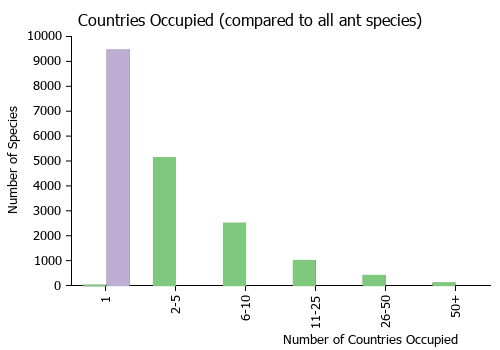
|
Estimated Abundance
| Relative abundance based on number of AntMaps records per species (this species within the purple bar). Fewer records (to the left) indicates a less abundant/encountered species while more records (to the right) indicates more abundant/encountered species. |

|
Biology
Castes
.png) X-ray micro-CT scan 3D model of Zasphinctus lumumbai (worker) prepared by the Economo lab at OIST.
X-ray micro-CT scan 3D model of Zasphinctus lumumbai (worker) prepared by the Economo lab at OIST.
holotype See on Sketchfab. See list of 3D images.
Nomenclature
The following information is derived from Barry Bolton's Online Catalogue of the Ants of the World.
- lumumbai. Zasphinctus lumumbai Hita Garcia & Gómez, 2025: 29, figs. 3D, 4D, 5D, 6D, 7D, 8D, 10D, 11D, 12D, 13D, 14D, 15D, 16D, 17D, 18D, 19D, 20D, 21D, 22 (w.) DEMOCRATIC REPUBLIC OF THE CONGO.
Type Material
- Holotype: Pinned worker, Democratic Republic of Congo, Equateur, Mabali, Tsuhapa River (Bikoro Terr.), Foret Inondée, Humus, collection code ANTC39356, IX.1959 (N. Leleup) (MRAC: MRACFOR0010007). [specimen re-mounted by KGA 2022]