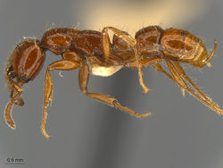
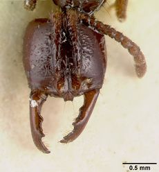
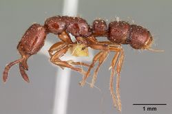
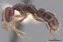
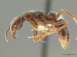
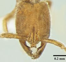
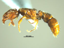
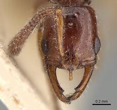
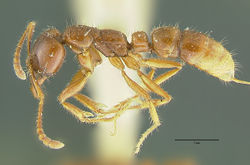
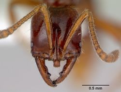
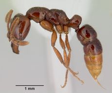
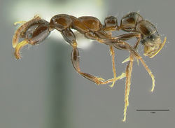
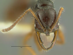
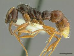
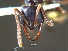
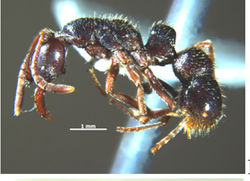
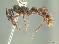
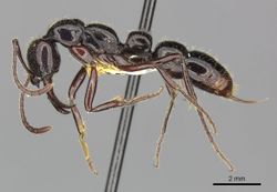
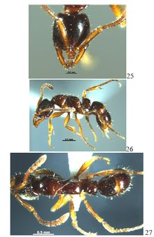
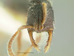
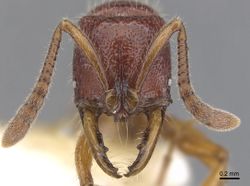
Key to Myopias workers
This worker key is based on: Probst, R.S., Guenard, B. and Boudinot, B.E. 2015. Toward understanding the predatory ant genus Myopias (Formicidae: Ponerinae), including a key to global species, male-based generic diagnosis, and new species description. Sociobiology. 62:192-212. doi:10.13102/sociobiology.v62i2.192-212
The key presented below represents the first attempt to provide a global worker-based identification resource to the species of Myopias. Mandibular morphology was found the most valuable, if not the most challenging character for which to describe patterns given the remarkable diversity in mandibular form. Second to the mandibles, form of the median clypeal lobe was highly informative.
Notes: Absolute characters are indicated by a full stop; these characters are sufficient alone to separate the particular taxon from the others being contrasted. Polythetic characters for which multiple conditions must be met are separated by semi-colons (consider them as boldface “and” conditional statements). Two Bill Brown morphospecies are difficult to key, but may be distinguished from described species as follows: M. axinipelta has a remarkably large median clypeal lobe, with strongly anteriorly-divergent lateral margins and a distinctly-margined wedge-shaped dorsal face; M. lacunosa has an almost perfectly rectangular head, with completely parallel, linear lateral margins, coarse lacunate sculpture, and very short, robust scapes. The following species were excluded: M. kuehni, a gyne-based species; M. papua and M. philippinensis, as they cannot be confidently placed in the key; and the subspecies M. bidens polita and M. maligna punctigera. Additionally, species related to M. tenuis are the least likely to key successfully, as there are new species to be described and valid taxa need re-evaluation. Regard this as a heuristic, coarse, preliminary key that requires testing with a broader intraspecific sampling and that will benefit from more refined character evaluation.
You may also be interested in Myopias
1
- Mandible elongate; in full-face view lateral margin convex; prebasal tooth strongly produced medially, subtended by short masticatory margin blade which diverges medially relative to lateral margin, and followed by strong concavity (Fig 7A) . . . . . Myopias concava
- Mandible elongate or short; in full-face view lateral margin convex or linear; prebasal tooth not strongly produced medially, subtended by short or long masticatory margin blade which may or may not be divergent relative to lateral margin, and not followed by strong concavity . . . . . 2
2
return to couplet #1
- Masticatory mandibular margin with conspicuously produced basal angle followed by a distinct concavity; masticatory margin blade between basal angle and prebasal tooth subequal in length to mandibular inner margin (Fig 7B). Median clypeal lobe apical margin concave, apicolateral corners conspicuously dentate (Fig 7B) . . . . . 3
- Masticatory mandibular margin usually without, but sometimes with conspicuously produced basal angle, if basal angle somewhat produced, then margin following linear to convex; masticatory margin blade between basal angle and prebasal tooth usually conspicuously shorter or longer than mandibular inner margin. Median clypeal lobe apical margin concave to convex, apicolateral corners rounded to angled . . . . . 7
3
return to couplet #2
- Compound eye well-developed; maximum diameter longer than malar space length (Fig 7C) . . . . . 4
- Compound eye reduced; maximum diameter shorter than malar space length (Fig 7D) . . . . . 5
4
return to couplet #3
- Median clypeal lobe length subequal to width in full-face view (Fig 7E) . . . . . Myopias bidens
- Median clypeal lobe length considerably less than width in full-face view (Fig 7F) . . . . . Myopias breviloba
5
return to couplet #3
- Anterior mesonotal margin raised well above posterior margin in profile view; mesonotal dorsum forming angle with propodeal dorsum . . . . . Myopias castaneicola
- Anterior mesonotal margin not or barely raised above posterior margin in profile view; mesonotal dorsum more-or-less continuous with propodeal dorsum, excluding metanotal impression . . . . . 6
6
return to couplet #5
- Median clypeal lobe comparatively large; anterolateral denticles pronounced. Scapes not reaching posterior head margin in full-face view. Lateral head margins weakly convex (Fig 7G) . . . . . Myopias mayri
- Median clypeal lobe comparatively small; anterolateral denticles weak. Scapes surpassing posterior head margin in full-face view. Lateral head margins strongly convex (Fig 7H) . . . . . Myopias trumani
7
return to couplet #2
- Median clypeal lobe absent or strongly reduced and without distinct apicolateral corners . . . . . 8
- Median clypeal lobe present, with or without distinct apicolateral corners . . . . . 10
8
return to couplet #7
- Mandible length in full-face view about equal to half head width. Prebasal tooth extremely robust, larger than compound eye . . . . . Myopias amblyops
- Mandible length in full-face view greater than half head width. Prebasal tooth not robust, smaller than compound eye . . . . . 9
9
return to couplet #8
- Prebasal tooth recurved in full-face view. With head in profile view, basal half of masticatory margin strongly produced anterodorsally, forming spectacular lobe . . . . . Myopias lobosa
- Prebasal tooth directed medially in full-face view. With head in profile view, basal half of masticatory margin not produced anterodorsally . . . . . Myopias cribriceps
10
return to couplet #7
- Head, mesosoma, and metasoma nearly completely covered with fine sculpture, rendering cuticle somewhat opaque (Fig 8A) [note: a Bill Brown morphospecies, “blanadelphe”, keys here; distinguishing characters are uncertain] . . . . . 11
- Head, mesosoma, and metasoma not covered with very dense fine sculpture; cuticle predominantly shining, even if sculpture present (Fig 8B) . . . . . 13
11
return to couplet #10
- Petiolar sternum bidentate; subpetiolar process followed by second triangular process. Compound eyes absent . . . . . Myopias nops
- Petiolar sternum unidentate; subpetiolar process not followed by second triangular process. Compound eyes represented by single pin-prick-like facets . . . . . 12
12
return to couplet #11
- Posterior head margin angularly concave in full-face view. Lateral margins of median clypeal lobe parallel. Dorsal face of petiole convex in profile view . . . . . Myopias shivalikensis
- Posterior head margin more-or-less linear in full-face view, but not angularly concave. Lateral margins of median clypeal lobe converging apically. Dorsal face of petiole linear in profile view . . . . . Myopias menba
13
return to couplet #10
- Mandible somewhat (Fig 8C) to extremely elongate (Fig 8D) and linear in full-face view; lateral margin linear and parallel or slightly subparallel with medial margin for most of mandibular length; basal mandibular margin considerably shorter than inner mandibular margin; basal mandibular margin oriented more-or-less subparallel with chord length of mandible, thus basal mandibular blade narrow [note: a Bill Brown morphospecies, “pisinna”, keys here, and may be differentiated from subsequent species by the honey-yellow coloration, short scapes, and mandibular form] . . . . . 14
- Mandible neither elongate nor linear in full-face view; lateral margin convex and not parallel with medial margin; basal mandibular margin well-developed, longer than inner mandibular margin; basal mandibular margin divergent from chord-length of mandible, thus basal mandibular blade broad . . . . . 31
14
return to couplet #13
- Head smooth, with or without dilute weak punctae. Median clypeal lobe lateral margins parallel to weakly diverging apically . . . . . 15
- Head with distinct, fine-to-coarse, conspicuous punctures or foveae, or head striate. Median clypeal lobe lateral margins parallel, converging apically or strongly diverging apically [note: a Bill Brown morphospecies, “epeirica”, keys here, and may be differentiated from the following species by presence of dense pubescence on the head, legs, and antennae] . . . . . 23
15
return to couplet #14
- Median clypeal lobe about three times as long as broad [note: placed based on description in Emery (1900)] . . . . . Myopias xiphias
- Median clypeal lobe at most twice as long as broad . . . . . 16
16
return to couplet #15
- Frontal lobes expanded, maximum lateromedial width almost twice anteroposterior length of median clypeal process. Basal blade of mandibular masticatory margin from basal angle to base of prebasal tooth shorter than basal mandibular margin. Prebasal tooth robust, length slightly less than half width of basal blade. Subpetiolar process wedge-shaped, anteroposteriorly broad and dorsoventrally tall in profile view. Compound eyes reduced (~ 5 facets); antennal scapes robust and short, not reaching posterior head margin in full-face view . . . . . Myopias darioi
- Frontal lobes not as expanded, maximum width less twice median clypeal lobe length, although may be about as broad as lobe. Basal blade of mandibular masticatory margin longer than basal mandibular margin. Prebasal tooth fine or robust, but considerably shorter than basal blade length. Subpetiolar process not as above: fine and subrectangular or somewhat broad and anteroventrally truncate, dorsoventrally short. Compound eyes reduced, represented by one to several facets, or eyes large ( > 7 facets); antennal scapes robust or slender, reaching posterior head margin or not . . . . . 17
17
return to couplet #16
- Propodeum weakly to strongly convex in profile view; meeting mesonotum at distinct angle . . . . . 18
- Propodeum sublinear in profile view; more-or-less continuous with mesonotum . . . . . 19
18
return to couplet #17
- Compound eye composed of over 20 ommatidia. Basal blade of mandibular masticatory margin subequal to length of basal mandibular margin. Propodeal dorsum smoothly convex in profile view . . . . . Myopias latinoda
- Compound eye composed of less than 20 ommatidia. Basal blade of mandibular masticatory margin considerably longer than basal mandibular margin. Propodeal dorsum unevenly convex in profile view . . . . . Myopias chapmani
19
return to couplet #17
- Compound eyes small, with less than 10 ommatidia . . . . . 20
- Compound eyes large, with over 10 ommatidia [note: placement here based on original descriptions in Crawley (1924) and Emery (1901); supposedly these two species may be distinguished by scape length, with M. crawleyi having scapes which barely fail to reach the posterior head margin and M. levigata having scapes which slightly exceed the posterior head margin] . . . . . Myopias crawleyi . . . Myopias levigata
20
return to couplet #19
- Compound eye composed of ≥ 4 ommatidia . . . . . Myopias emeryi
- Compound eye composed of ≤ 3 ommatidia [note: M. santschii Viehmeyer, 1914 keys here based on the original description, but cannot be confidently placed further] . . . . . 21
21
return to couplet #20
- Scapes failing to reach to reaching posterior head margin when laid back in full-face view. Small; head width < 0.70 mm . . . . . Myopias tenuis
- Scapes overreaching posterior head margin when laid back in full-face view. Larger; head width > 0.90 mm . . . . . 22
22
return to couplet #21
- Prebasal tooth situated at about mandibular midlength. Head capsule nearly broad as long (HW/HL*100 = 99). Mandibles robust . . . . . Myopias media
- Prebasal tooth situated at about distal third of mandible. Head capsule longer than broad (HW/HL*100 ~ 90). Mandibles thin . . . . . Myopias julivora
23
return to couplet #14
- Median clypeal lobe lateral margins strongly divergent apically, meeting apical margin at pronounced angles (Fig 8E) . . . . . 24
- Median clypeal lobe lateral margins parallel, convergent, or weakly divergent apically, meeting apical margin at rounded angles . . . . . 27
24
return to couplet #23
- Abdominal terga III and IV smooth and shining . . . . . 25
- Abdominal terga III and IV foveate . . . . . 26
25
return to couplet #24
- Metanotal impression conspicuous. Smaller; head width < 1 mm. Scapes robust; maximum width longer than malar space length . . . . . Myopias maligna
- Metanotal impression weak, inconspicuous. Larger; head width > 1 mm. Scapes slender; maximum width shorter than to subequal to malar space length . . . . . Myopias hollandi
26
return to couplet #24
- Scapes long, reaching or surpassing posterior head margin when laid back in full-face view. Petiolar sternum linear posterior to anteroventral tooth in profile view. Petiolar node width about equal to length in dorsal view . . . . . Myopias conicara
- Scapes shorter, not reaching posterior head margin when laid back in full-face view. Petiolar sternum convex posterior to anteroventral tooth in profile view. Petiolar node broader than long in dorsal view . . . . . Myopias hania
27
return to couplet #23
- Median clypeal lobe apical margin medially notched. Anterior mesonotal margin raised well above posterior margin. Propodeal dorsum strongly convex in profile view . . . . . 28
- Median clypeal lobe apical margin linear to weakly convex. Anterior mesonotal margin not or barely raised above posterior margin. Propodeal dorsum more-or-less linear in profile view . . . . . 29
28
return to couplet #27
- Notch of median clypeal lobe apical margin shallow. Median clypeal lobe shorter, broader, and tapering to apex . . . . . Myopias gigas
- Notch of median clypeal lobe deep. Median clypeal lobe longer, narrower, with sides subparallel . . . . . Myopias loriai
29
return to couplet #27
- Median clypeal lobe very short; lateral margins parallel. China . . . . . Myopias luoba
- Median clypeal lobe longer; lateral margins parallel to weakly diverging apically. New Guinea or Australia . . . . . 30
30
return to couplet #29
- Petiolar node lateral margins sublinear, conspicuously diverging posteriorly in dorsal view. Posteroventral angle of subpetiolar process acute, sharp. New Guinea . . . . . Myopias ruthae
- Petiolar node lateral margins convex, subparallel to weakly diverging posteriorly in dorsal view. Posteroventral angle of subpetiolar process obtuse, weakly rounded. Australia . . . . . Myopias densesticta
31
return to couplet #13
- Median clypeal lobe tridentate, with apicomedial process in addition to apicolateral corners (Fig 8F) . . . . . 32
- Median clypeal lobe bidentate, without apicomedial process in addition to apicolateral corners . . . . . 33
32
return to couplet #31
- Apicomedial process of median clypeal lobe produced anteriorly. Basal blade of mandibular masticatory margin weakly produced medially [note: placement based on description in Crawley (1924)] . . . . . Myopias modiglianii
- Apicomedial process of median clypeal lobe not produced anteriorly. Basal blade of mandibular masticatory margin conspicuously produced medially . . . . . Myopias mandibularis
33
return to couplet #31
- Basal angle of mandibular masticatory margin dentate (Fig 8G). Mandibles very broadly triangular in full-face view . . . . . Myopias delta
- Basal angle of mandibular masticatory margin edentate (Fig 8H). Mandibles less broadly triangular in full-face view . . . . . 34
34
return to couplet #33
- Basal angle of mandible distinct. Median clypeal lobe length and width subequal. China . . . . . Myopias daia
- Basal angle of mandible indistinct, rounded. Median clypeal lobe broader than long. Australia . . . . . Myopias tasmaniensis


