File:Billen and Verbesselt 2016b F2.jpg

Original file (1,700 × 2,057 pixels, file size: 1.24 MB, MIME type: image/jpeg)
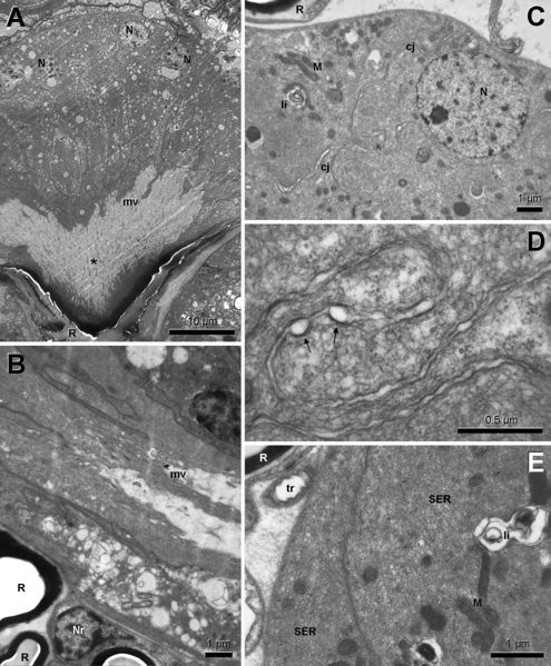
Figure 2. Electron micrographs of anterior part of Pavan’s gland in minor worker. A. Low magnification view in cross section showing columnar cells with basally located nuclei (N), irregular apical border and large subcuticular space (asterisk). B. Apical cytoplasm with irregular microvilli (mv) and reservoir all (R); Nr: nucleus of reservoir wall cell. C. Basal cytoplasm with round nuclei, lamellar inclusions (li), numerous mitochondria (M) and contorted cell junctions (cj). D. Detail of contorted cell junction in lower epithelial region with ocally round intercellular spaces (arrows). E. Detail of basal cytoplasm with well-developed smooth endoplasmic reticulum (SER) and numerous mitochondria. tr: tracheole.
Billen, J. and S. Verbesselt. 2016. Morphology and ultrastructure of Pavan’s gland of Aneuretus simoni (Formicidae, Aneuretinae). Asian Myrmecology. 8:101-106. doi:10.20362/am.008017
File history
Click on a date/time to view the file as it appeared at that time.
| Date/Time | Thumbnail | Dimensions | User | Comment | |
|---|---|---|---|---|---|
| current | 01:49, 28 November 2017 |  | 1,700 × 2,057 (1.24 MB) | Lubertazzi (talk | contribs) | Figure 2. Electron micrographs of anterior part of Pavan’s gland in minor worker. A. Low magnification view in cross section showing columnar cells with basally located nuclei (N), irregular apical border and large subcuticular space (asterisk). B. A... |
You cannot overwrite this file.
File usage
The following page uses this file: